
Spatial Omics and Single-Cell Biology: Mapping the Next Generation of Precision Therapeutics
Today, we examine how spatial omics and single-cell biology are overcoming the limitations of bulk sequencing by mapping gene activity directly within intact tissue architecture. Discover how this architectural shift is accelerating target discovery, refining biomarker validation, and powering the next generation of precision therapeutics.
This week in the Guardrail… we examine how spatial omics and single-cell biology are overcoming the limitations of bulk sequencing by mapping gene activity directly within intact tissue architecture. Discover how this architectural shift is accelerating target discovery, refining biomarker validation, and powering the next generation of precision therapeutics.
By Michael Bronfman
August 17, 2026
Traditional genomic and transcriptomic analyses have long served as the foundation of biomedical research, yet they carry a fundamental structural limitation. Standard sequencing protocols require researchers to grind up heterogeneous tissue samples to extract their DNA or RNA. While this bulk extraction yields valuable genetic readouts, it completely destroys the spatial architecture of the tissue sample. The homogenization process blends thousands or millions of distinct cells into a single average measurement. This leaves researchers unable to observe how individual cell types were originally organized, how they communicated through local signaling, or how they interacted within their microenvironment.
Spatial transcriptomics and single-cell biology directly overcome this limitation. By enabling scientists to profile gene activity directly within an intact tissue section, these combined methodologies preserve critical spatial coordinates alongside comprehensive molecular signatures. By bridging single-cell sequencing with high-resolution optical imaging, spatial omics maps the exact location of individual cell populations while detailing their corresponding gene expression profiles inside native tissue architecture.
This technical shift is accelerating preclinical research, refining target discovery, and transforming drug development strategies across major therapeutic areas.
The Technological Shift: Moving Beyond Bulk Averages
For decades, drug discovery teams relied heavily on bulk sequencing data to identify therapeutic targets. Bulk measurements, however, routinely mask low-abundance cell populations or localized transcriptional shifts. A candidate drug target that appears minimally expressed across an entire tumor lysate may actually be highly expressed within a localized cluster of aggressive cells at the invasive tumor margin.
Single-cell RNA sequencing introduced higher resolution by isolating individual cells prior to library preparation, enabling researchers to classify distinct cell subtypes and rare phenotypes. Yet, the physical dissociation step required for single-cell RNA sequencing severs extracellular matrix contacts and destroys spatial organization.
Spatial omics platforms bridge this remaining gap. By utilizing spatially barcoded solid-phase capture arrays or multiplexed in situ hybridization, these technologies capture mRNA transcripts directly from intact tissue sections. 1
Transformative Impact Across Key Therapeutic Areas
Integrating spatial profiling into preclinical drug development pipelines provides unprecedented clarity into complex human diseases, directly improving target validation and risk assessment.
Oncology and the Tumor Microenvironment
Cancerous tissue is exceptionally complex, consisting of malignant subclones, infiltrating immune cells, stromal fibroblasts, blood vessels, and dense extracellular matrix components. Spatial omics allows oncology researchers to deconstruct the tumor microenvironment with high spatial resolution.
Immune Exclusion Zones: Spatial profiling explains why checkpoint inhibitors succeed in certain regions of a tumor while failing in others. Researchers can identify physically restricted immune cells trapped within dense fibrotic borders, which prevent them from penetrating the core tumor tissue.
Therapeutic Resistance Niches: Tumor regions located near hypovascularized or hypoxic cores display distinct stress-response signatures. Mapping these microdomains clarifies how localized environments shield malignant subclones from systemically administered therapies.
Tertiary Lymphoid Structures: Identifying the presence, cellular composition, and spatial layout of tertiary lymphoid structures within solid tumors offers strong predictive value for immunotherapy response. This enables clinical teams to stratify patient cohorts more effectively during early clinical trials.
Neuroscience and Structural Brain Mapping
The central nervous system depends entirely on an organized spatial architecture, in which local neuronal circuits govern functional signals. Bulk sequencing of brain tissue offers limited actionable insight into circuit-level pathologies.
Neuronal Circuit Profiling: Spatial transcriptomics maps the complex laminar organization of the cerebral cortex, charting gene signatures across distinct cortical layers and subcortical structures.
Neurodegenerative Pathology Progression: In conditions such as Alzheimer disease or Parkinson disease, pathological features such as amyloid plaques and neurofibrillary tangles form in localized regions. Spatial profiling allows researchers to evaluate transcriptomic shifts occurring in microglial cells and neurons immediately adjacent to these protein aggregates, comparing them directly to distant healthy tissue.
Blood-Brain Barrier Integrity: Mapping the vascular parenchymal interface helps researchers evaluate how local inflammatory signals compromise active transport across the blood-brain barrier. This aids the rational design of central nervous system drug delivery vehicles.
Immunology and Inflammatory Pathology
Immune responses depend on coordinated cellular migration and localized paracrine signaling within specialized tissue structures.
Lymph Node Architecture: Spatial methods map how immune cell subsets, including dendritic cells, helper T cells, and cytotoxic lymphocytes, reorganize within follicular and paracortical regions during antigen presentation or autoimmune activation.
Autoimmune Niches: In chronic inflammatory conditions such as rheumatoid arthritis or Crohn disease, spatial omics identifies local cellular niches that drive tissue damage. This uncovers localized cytokine networks that systemic blood profiling fails to detect.
Integrating Spatial Omics into the Drug Discovery Pipeline
Pharmaceutical R&D organizations are transitioning from bulk tissue profiling to spatially resolved experimental workflows. This workflow integration enhances multiple stages of early drug development.
Target Identification and Validation
By overlaying high-resolution transcript profiling onto tissue histology, discovery teams can verify whether a target is expressed specifically within disease-driving cells or broadly across healthy tissue. Target evaluation at the tissue microdomain level reduces off-target toxicity risks early in discovery.2
Pharmacodynamics and Tissue Distribution
Assessing candidate drug efficacy requires confirming that a therapeutic agent reaches target cells at effective concentrations within complex tissue. Combining spatial transcriptomics with mass spectrometry imaging or spatial proteomics allows research teams to map drug molecule concentration alongside downstream gene expression changes in the exact same tissue section.
Biomarker Discovery for Clinical Stratification
Phase 2 and Phase 3 clinical trial failures frequently stem from unaddressed patient heterogeneity. Analyzing intact patient biopsy tissue reveals spatial biomarkers, such as cell-to-cell proximity scores or immune infiltration indices, that identify patient subgroups most likely to achieve clinical response.3
Comparative Analysis of Primary Spatial Omics Technologies
Modern spatial biology platforms fall into two main technical categories: sequencing-based spatial capture arrays and imaging-based in situ hybridization technologies. Selecting the appropriate technology depends on research goals, resolution requirements, and target gene panel depth.Sequencing-based capture platforms, such as array-based transcript mapping systems, provide unbiased, high-throughput coverage across the entire genome. These systems are ideal for exploratory target discovery where relevant biological pathways are not yet fully defined.
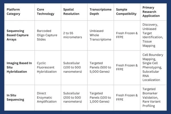
In contrast, imaging based platforms achieve subcellular resolution. These systems utilize targeted fluorescent probe panels to detect individual mRNA molecules within defined cell boundaries. 4
Laboratory Adoption Considerations and Technical Protocols
Successfully establishing a spatial transcriptomics workflow within a biomedical laboratory requires addressing sample preservation, assay selection, and computational data processing infrastructure.
Sample Preparation: FFPE vs Fresh Frozen Tissue
Tissue sample preparation remains a critical variable influencing spatial assay performance.
Fresh Frozen (FF) Tissue: Preserves high-quality RNA integrity, making it optimal for whole transcriptome sequencing-based profiling. However, collecting and preserving fresh frozen samples requires continuous cold-chain storage, which can be difficult to maintain across multisite clinical trial networks.
Formalin Fixed Paraffin Embedded (FFPE) Tissue: Represents the standard preservation format for historical clinical pathology archives. While formalin fixation induces crosslinking and RNA degradation over time, modern probe-based capture chemistries allow robust RNA transcript detection directly from archival FFPE tissue blocks.
Resolution and Multiplexing Tradeoffs
Laboratories must balance transcriptomic breadth against spatial resolution based on specific experimental requirements.
Whole Transcriptome Array Profiling: Measures overall gene expression across thousands of mRNA species across array capture spots. While comprehensive, spatial resolution on standard arrays may encompass multiple adjacent cells per spot, requiring bioinformatic deconvolution to estimate individual cell contributions.
Subcellular Single-Molecule Imaging: Uses targeted, multiplexed probes to achieve resolution down to the hundreds of nanometers. These methods allow direct visualization of intracellular transcript localization within specific organelles or near cell membranes, but require preselecting specific gene-target panels.
Computational Pipelines and Data Scale
Spatial transcriptomics experiments produce extensive datasets combining high-resolution multi-channel image stacks with dense molecular expression matrices. Processing these datasets requires specialized bioinformatic tools capable of handling image registration, cell boundary segmentation, background signal suppression, and spatial neighborhood clustering.
Market Trajectory and Commercial Dynamics
Driven by demand for targeted therapies and precision oncology, the spatial biology market is growing rapidly. Commercial market reports project strong expansion across reagents, instruments, and analytical software through the coming decade.
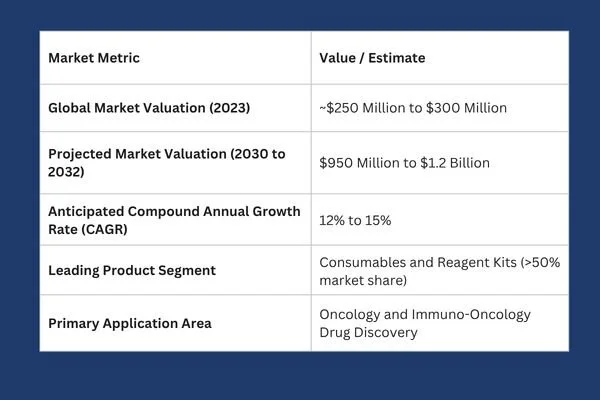
Key drivers powering this commercial expansion include automated tissue-processing instruments, decreasing sequencing costs, improved single-cell segmentation algorithms, and expanding spatial foundation models for automated pathology analysis.
Future Directions: Multimodal Spatial Omics and Artificial Intelligence
The field of spatial biology is rapidly moving beyond single-analyte transcript mapping toward integrated multimodal measurements. Modern experimental platforms enable concurrent spatial profiling of transcripts, functional proteins, epigenetic chromatin accessibility, and small-molecule metabolites within the exact same tissue section.
Concurrently, artificial intelligence models and spatial foundation frameworks are reshaping tissue analytics. Advanced machine learning algorithms can now correlate standard hematoxylin and eosin (H&E) pathology images with underlying spatial gene expression patterns. By training neural networks on matched spatial transcriptomic datasets, researchers can predict localized molecular signatures directly from routine histological slides, unlocking deep biological insight from archival clinical pathology banks.
As these technologies continue to mature and protocol costs decline, spatially resolved biology will evolve from an exploratory research tool into a mandatory standard across pharmaceutical target validation, safety testing, and translational medicine. By preserving the architectural landscape of human tissue, spatial omics is establishing a more accurate foundation for precision drug development.
1 National Library of Medicine, Zhai, Chen & Deng. Researchers can view both the identity of the cell and its exact cellular neighborhood, linking transcriptomic expression directly to tissue histology. pmc.ncbi.nlm.nig.gov
2 Illumina, "What is transcriptomics? A complete guide to gene expression analysis, July 29, 2026. Comprehensive guides on transcriptomic workflows can be found via Illumina transcriptomics overview. illumina.com
3 Atlantis Bioscience, Discovering Solutions for Translational Science. Researchers seeking technical validation tools, reagents, and spatial profiling solutions can explore resources hosted at Atlantis Bioscience. atlantisbioscience.com
4 University of Wisconsin-Madison, Biotechnology Center Gene Expression Core. 10xGenomics Xenium in Situ Spatial Platform. Specialized commercial platforms in this space include the 10x Genomics Xenium in situ platform. biotech.wisc.edu S and the Vizgen MERSCOPE platform. vizgen.com
Our expert advisors accelerate your therapeutic candidates from target validation to clinical triumph. Contact Metis Consulting Services today to integrate cutting-edge spatial transcriptomics and precision workflow strategies into your drug discovery pipeline.
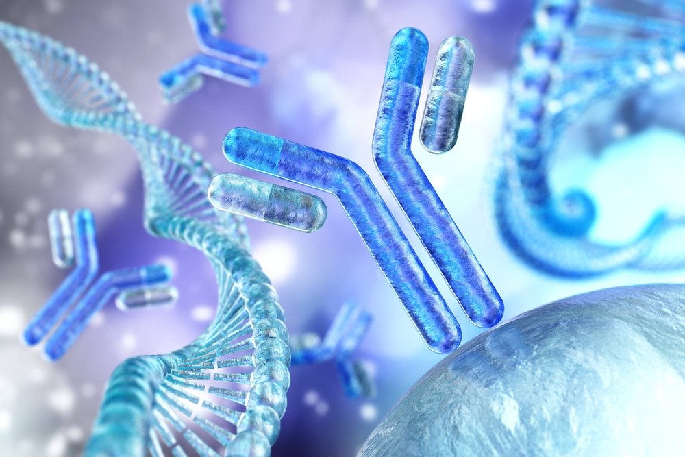
The Rise of Computer-Designed Antibodies and What It Means for Therapeutics
Recently, researchers have developed advanced computational tools to design antibodies in ways previously considered unattainable. This development could accelerate the discovery of new treatments and expand the range of treatable diseases.
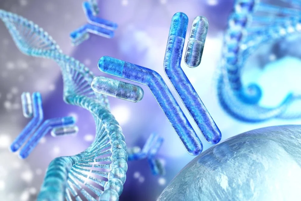
As the field of drug discovery undergoes a monumental shift toward computational efficiency, staying ahead of regulatory and quality benchmarks is essential for success. This week in the Guardrail, we examine how the rise of computer-designed antibodies is redefining what is possible for modern therapeutics
By Michael Bronfman for Metis Consulting Services
January 12, 2026
Life sciences are undergoing a significant shift in drug discovery. Historical methods depend on animal testing, extensive molecular libraries, and lengthy trial-and-error cycles. Recently, researchers have developed advanced computational tools to design antibodies in ways previously considered unattainable. This development could accelerate the discovery of new treatments and expand the range of treatable diseases.
An antibody is a type of protein that your immune system makes to recognize and bind to substances such as viruses or bacteria. Because antibodies can bind very specifically to targets, they make excellent therapeutic drugs. There are already many antibody medicines on the market for cancer, autoimmune diseases, and infectious diseases. But creating new antibody drugs with traditional methods is slow, costly, and often unpredictable. Researchers now use advanced computer models to guide antibody design. These models “learn” from large-scale biological datasets to propose new proteins that bind targets with high precision and affinity.
Learn more at the PubMed review on antibody design advances.
In this article, we will explore why computer-designed antibodies are now possible, how they may improve treatments, and what challenges remain.
What Are Antibodies and Why Are They Important
Antibodies are proteins produced by the immune system to recognize and attach to antigens. They look like a “Y” shape with two arms that grab the target. The part of the target that binds the ligand is called the binding region. This region can be fine-tuned to stick firmly to one specific target protein. Many modern drugs are antibodies because they can block harmful proteins without interfering with normal body functions.
More than one hundred therapeutic antibodies have been approved for use in humans. Some of these work by blocking signals that promote cancer-cell growth. Others mark infected cells so the immune system can destroy them. Because these drugs are specific, they often cause fewer side effects than traditional small-molecule drugs. But making new antibody drugs requires many months of lab work. Traditional methods involve immunizing animals or screening large collections of molecules to identify rare, suitable candidates. These processes are expensive and sometimes fail to find suitable matches for challenging targets.
How Computers Can Help Design Antibodies
Computational design changes this process by using large datasets and predictive models to propose antibody candidates before they are made in the lab. These tools examine protein shapes and their interactions. They can then suggest novel antibody sequences that should fold into shapes that bind strongly to a chosen target.
One significant advance came when research groups taught computers to build antibodies from scratch. At the University of Washington, scientists created complete antibodies entirely on computers. They controlled where the antibody would bind and then tested the designs in the lab. Many of these computer designs folded and bound targets as expected. The result suggests computers can help design new antibody drugs much faster than traditional methods.
In addition to full antibody design, other computational tools can optimize specific antibody regions. For example, they can predict how changes in amino acid sequence might increase binding strength or reduce unwanted interactions. The combination of prediction and testing accelerates the full path from idea to experimental candidate.
Examples of Progress in Therapeutic Antibody Design
A recent milestone in this field is Imneskibart (AU-007). This is the first fully computer-designed antibody to enter clinical trials. It was created to bind a specific part of the immune system and modulate immune responses in cancer without causing the common toxic side effects seen with older therapies. The fact that this medicine has reached clinical testing is significant proof of concept for computational design methods.
Another example is in the Reuter’s report on industry partnerships between a U.S. biotech company and a global pharmaceutical firm. They expanded their research collaboration to focus on protein and antibody design using advanced computational platforms. These platforms can propose designs and move them to initial laboratory testing in only a few weeks, compared to months or years with older methods.
Alongside specific antibody drugs, research groups worldwide are using technology to tackle challenging disease targets. That includes chronic infections, rapidly mutating cancer antigens, and proteins previously considered undruggable. These new tools give scientists more control over the design process and reduce reliance on random screening as shown in the Pharmaceutical Journal on the future of antibody drugs.
Benefits of Computer-Designed Antibodies
There are several essential benefits to designing antibodies with computational methods:
1. Speed: Historical discovery can take years. Computational design can quickly narrow down promising candidates and may cut months from early phases of drug discovery.
2. Precision: Computers can predict the exact spot, or epitope, on a target protein where an antibody will bind. This precision helps create drugs that block specific functions without interfering with other parts of the body.
3. Better screening: Instead of testing millions of random molecules, researchers can use computational filters to test just a few dozen promising candidates in the lab. This reduces cost and waste.
4. Hard targets: Some disease targets are very difficult to bind with traditional methods. Computational design can explore new molecular shapes that might succeed where older methods fail.
5. Reduced side effects: By designing antibodies that bind only to intended targets, there is a potential for fewer off-target interactions that cause adverse effects.
In many ways, these new computer-guided tools behave like powerful microscopes. They allow scientists to see and test possibilities that were once invisible or unreachable with older methods.
Learn more at the University of Washington’s report on computer-designed antibodies in nature.
Challenges and Limitations
Even though these new design methods are powerful, they are not yet perfect. A central challenge is validation. Computers can propose many candidate molecules, but only some of these actually fold and bind as predicted in real lab conditions. Researchers still need to test candidates experimentally before they become drug candidates.
Another challenge is that the design models depend on large datasets of known protein structures. If a target is very different from anything in the datasets, the design models may not make accurate predictions. Scientists are working to expand these training sets and improve model performance.
There are also development hurdles. Even after a good candidate is found, it must be manufactured reliably and safely. The pathway from an early design to an approved drug includes multiple steps, including tissue testing, toxicology studies, and clinical trials, which remain costly and time-consuming.
Finally, there is the question of accessibility. Currently, many of the most advanced design tools are available only to large companies or research institutions with significant computing resources. Making these tools more widely available could help smaller organizations contribute to discovery and innovation.
What This Means for Future Medicines
The rise of computer-designed antibodies may change what is possible in medicine. Because these tools speed up early discovery, they could bring new treatments to patients faster than ever before. This could be valuable for diseases that have no good treatments today.
For example, researchers are using computational design to pursue cancer targets that mutate rapidly and immune molecules with complex structures. These targets were once considered too difficult for standard methods. If computers can identify stable designs for these targets, new therapies could reach patients in need.
In addition, the improved precision may lead to safer medicines. With a better understanding of how an antibody binds its target, scientists can avoid unintended effects that cause harm. As computational tools improve and large datasets grow, the accuracy of these predictions will also increase.
Because of the faster pace of design, new antibody treatments could be developed for emerging infectious diseases. During a pandemic or outbreak, the ability to rapidly design antibodies that neutralize a threat could save many lives.
Overall, we are nearing a time when computers are normal parts of the drug discovery toolkit. They do not replace human scientists but give them powerful new tools to explore possibilities that would be very hard to test with old methods.
The growth of computer-designed antibodies shows how technology can reshape life sciences. These tools bring speed, precision, and new possibilities to therapeutic discovery. While challenges remain, the progress so far suggests a future where new treatments can be developed more rapidly and more safely. For patients with unmet medical needs, this change could be life-changing.
The promise of these methods comes from their ability to transform what used to be guesswork into guided design. As computational capabilities continue to improve and merge with experimental science, the pace of discovery will only increase. The future of therapeutics will include more medicines that were first conceived on a computer screen and then tested and refined in the lab.
As your organization adopts cutting-edge technologies like computational antibody design, Accelerate Innovation with Metis Consulting Services. Navigating the complexities of quality, regulatory strategy, and data management is more challenging than ever. We provide the expert guidance you need to transform these technological breakthroughs into safe, market-ready therapies. Contact Metis Consulting Services today to schedule a consultation and ensure your pipeline is built on a foundation of wisdom and precision. hello@metsconsultingservices