
This week in The Guardrail, explore how spatial omics and single-cell biology are replacing traditional, blended tissue analysis. Preserving the cellular microenvironment, researchers watch spatial interactions unfold in real time. Will patients get the benefits?
By Michael Bronfman
November 9, 2026
Mapping the human body used to be like studying a bustling city from a satellite in outer space. You could see that a city existed, but you had no idea what was happening on individual streets, inside family homes, or between next-door neighbors.
In medicine, researchers have spent decades trying to understand how our organs work and why deadly illnesses strike. For a long time, the standard method was bulk analysis. Lab scientists took diseased tissue, put it into a liquid solution, and ground it like fruit in a blender. Then, they measured all the proteins and genes floating inside that mixture.
While that smoothie approach gave researchers a broad list of parts, it destroyed the tissue's structure. If you blend apples, bananas, and strawberries together, you cannot tell where each individual fruit begins or which slices touched each other during the process.
Then single-cell biology arrived. This method let scientists break tissue apart gently so they could examine individual cells one by one. It was a massive step forward. Scientists could finally divide cells into clean groups like healthy cells, immune defenders, and diseased cells.
Even with that progress, pulling cells apart had a major flaw. When you remove a cell from its home tissue, you wipe away its address and its neighborhood. You lose the vital biological signals it passes to its neighbors every single second.
A new field called spatial omics is changing modern drug discovery. By merging high-power microscopic imaging with genetic reading tools, spatial omics lets researchers read the chemical messages inside individual cells while leaving the tissue completely intact. This gives drug hunters an exact “street map” of human tissue..
What Is Spatial Transcriptomics?
To understand this new world, we have to look inside the cell nucleus. Inside every cell in your body, DNA acts like an instruction manual. When a cell needs to do a job, it makes working copies of those DNA pages using molecules called messenger RNA, or mRNA. This step is gene expression.
Reading all the mRNA copies inside a cell is called transcriptomics.
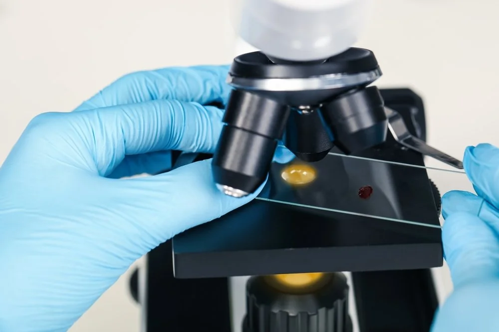
Spatial transcriptomics takes this reading technique onto real tissue. Instead of shaking cells free into plastic tubes, scientists mount a razor-thin slice of tissue onto a prepared slide. The slide's surface holds millions of capture spots. Each spot has a unique chemical tag that acts like a biological tracker.1
When the tissue sits on the slide, the mRNA molecules inside the cells stick directly to these capture spots. Advanced sequencing machines decode the genetic messages, while computers read the locator tags to pinpoint where each message came from.
Spatial transcriptomics lets researchers measure gene activity across an intact tissue slice. This reveals how cells communicate with their immediate neighbors. It is especially useful for unraveling solid tumors or mapping nerve pathways in the brain. Scientists no longer have to guess which cell sent a warning signal or which cell caught it. They can watch the physical “conversation” happen right before their eyes.
Spatial Transcriptomics Versus Spatial Omics
People sometimes use the words spatial transcriptomics and spatial omics like they mean the exact same thing, but they do not.
Spatial transcriptomics focuses on one thing: messenger RNA. It checks which genes are turned on and where those messages sit on the physical tissue grid.
Spatial omics is the larger family tree that includes spatial transcriptomics plus every other spatial measurement tool. Think of spatial transcriptomics as looking at only the written blueprints inside a building. Spatial omics looks at the blueprints, the steel framing, the electrical cables, the water pipes, and the waste bins all at once.
Within spatial omics, researchers track several biological layers across intact tissue:
- Spatial genomics maps DNA mutations and physical chromosome damage across different regions of an organ.
- Spatial proteomics tracks actual working proteins, cellular receptors, and structural fibers using tagged antibodies.2
- Spatial epigenomics studies the chemical tags that sit on top of DNA, showing which parts of the genetic code are unlocked or locked away.
- Spatial metabolomics identifies small molecules, nutrients, sugars, and fats to show how cells use energy across different tissue zones.
Spatial transcriptomics tells us what a cell plans to build. Spatial omics shows the full picture, from basic genetic instructions to real-time chemical energy.
Why The Microenvironment Matters
In human biology, location changes everything. Cells never work in total isolation. A cell acts one way when it touches a clear blood vessel, but it acts totally differently when trapped in deep inflammation or choked by scar tissue. We call this context the cellular microenvironment.
The microenvironment is crucial when fighting solid tumors. Cancer is rarely a simple clump of identical bad cells. A single tumor contains hyperactive cancer cells, dying cells starved of oxygen, fresh blood vessels feeding the mass, and immune cells sent by the body to kill the tumor.
Tumors survive by twisting their local environment to their advantage. They build thick walls of tough collagen protein that physically block immune cells from getting close. They also release confusing chemical signals that put killer T cells to sleep or trick them into ignoring the cancer entirely.
With spatial omics, scientists look directly into this combat zone. They see where immune cells crowd together, which ones break through the tumor perimeter, and which ones get stuck outside. This helps pharmaceutical teams design smarter cancer immunotherapies that guide the immune system through defensive shields.3
The same spatial rules govern the human brain. The brain contains billions of nerve cells linked through winding electrical networks. In conditions like Alzheimer disease or Parkinson disease, damage usually starts in tiny, distinct brain zones before spreading outward. Spatial tools let scientists see which specific nerve cells feel early stress, watch how resident immune cells called microglia react, and spot the exact moment a cellular pathway breaks down. 4.
The same spatial rules govern the human brain. The brain contains billions of nerve cells linked through winding electrical networks. In conditions like Alzheimer disease or Parkinson disease, damage usually starts in tiny, distinct brain zones before spreading outward. Spatial tools let scientists see which specific nerve cells feel early stress, watch how resident immune cells called microglia react, and spot the exact moment a cellular pathway breaks down.4
Turning Complex Biology Into Real Medicines
Bringing a brand new medicine to patients is one of the hardest tasks in modern science. It often takes over ten years and more than a billion dollars to move a medicine from an idea in a notebook to a bottle in a neighborhood pharmacy.
A main reason candidate drugs fail in human clinical trials is lack of efficacy. A chemical compound might kill cancer cells sitting flat on a plastic laboratory dish, but it fails completely inside a real 3-D human organ. Spatial omics helps solve this problem by giving drug makers realistic biological models during the earliest research stages:
- Finding Better Drug Targets: Scientists discover rare receptor proteins that show up only on diseased cells within specific tissue zones. This helps teams design medicines that attack disease while sparing nearby healthy organs.
- Verifying How Drugs Work: When testing a candidate molecule, researchers check whether the medicine reaches the center of a tumor and shuts down the target pathway it was designed to stop.
- Matching the Right Patient to the Right Drug: Sickness looks different in every patient. By studying tissue architecture in a small biopsy sample, doctors can tell whether a patient will respond to a specific immunotherapy or needs a completely different therapy right away.
- Checking Organ Safety Early: Spatial profiling reveals whether a candidate drug causes unwanted swelling or cell death in delicate bystander tissues, such as the kidneys or liver, long before human volunteers ever swallow a pill.5
The future of medicine is fundamentally spatial. The days of treating complex illnesses with blunt tools and identical treatments for every person are fading. By building visual maps of cellular activity, spatial biology is turning pharmaceutical development into a more exact, targeted science.
Moving From Raw Laboratory Data To Approved Cures
Gathering deep spatial data is only half the battle. A modern life sciences company can quickly generate billions of data points across hundreds of tissue slides. Yet raw image files and genetic tables cannot cure an ill patient on their own.
Research teams must organize these massive datasets, interpret the results through clinical eyes, run safe patient trials, and satisfy global health agencies like the United States Food and Drug Administration and the European Medicines Agency.
This journey demands strong partnerships. Specialized regulatory and data advisors help biotechnology teams build solid foundations for their discoveries:
Building Sound Regulatory Plans: Brand new technologies like spatial omics produce novel forms of biological evidence. Expert advisors show research teams how to present spatial biomarker data clearly so that trial designs meet formal submission rules at major health agencies.
Managing Complex Clinical Data: Spatial trials produce massive files that link high-resolution microscope photos with deep genetic code files. Modern data managers construct secure digital systems that protect data quality, organize patient records, and keep multi-center studies running smoothly.
Meeting Quality and Laboratory Rules: Moving a drug candidate into human testing means research labs must follow strict Good Laboratory Practice and Good Clinical Practice standards. Independent quality checks and clear laboratory procedures keep research accurate and compliant.
Applying Artificial Intelligence Responsibly: Modern spatial platforms depend on computer algorithms to identify cell types and predict cellular interactions. Quality teams review these algorithms to ensure computational models remain transparent, fair, and backed by clear biological evidence.
Protecting Patient Safety: As soon as an experimental drug enters clinical trials, tracking patient well-being becomes the top priority. Clinical safety teams monitor trial volunteers, catch negative side effects quickly, and protect patient safety through every clinical phase.
When careful regulatory guidance supports advanced spatial discoveries, life-saving medicines move out of laboratory freezers and into clinic rooms much faster. We help you bridge the gap between complex life science innovations and global regulatory compliance. Contact Metis Consulting Services today.
RESOURCES
