

Direct Cellular Engineering
Direct cellular engineering

This week in the Guardrail, we explore how shifting cellular engineering directly inside the human body could eliminate costly manufacturing delays and revolutionize patient access to life-saving treatments. Are you ready to overcome regulatory and delivery hurdles and bring next-generation cell therapies to market faster? Read on.
By Michael Bronfman
August 24, 2026
Traditional cell therapies have achieved extraordinary accomplishments in modern oncology. Chimeric antigen receptor T cell treatments have altered the prognosis for patients suffering from aggressive hematological malignancies like relapsed leukemias, lymphomas, and multiple myeloma. Despite these clinical achievements, the logistical structure of modern cell therapy remains fundamentally constrained.
The current standard of care relies entirely on an autologous ex vivo manufacturing paradigm. Clinicians must harvest T cells from a patient through leukapheresis, transport those living cells to dedicated Good Manufacturing Practice facilities, genetically reprogram them using viral vectors, expand them over several weeks, and deliver them back to the clinical site for reinfusion.
Manufacturing schedules routinely span three to five weeks. This process presents severe bottlenecks. Throughout this waiting window, rapidly progressing disease can cause patient deterioration or mortality before the engineered cells arrive. Intensive labor, stringent quality control, and specialized infrastructure drive treatment prices to hundreds of thousands of dollars per patient, excluding ancillary medical costs for lymphodepleting chemotherapy and prolonged hospital stays.
The Ex Vivo Bottleneck vs The In Vivo Solution
To understand the revolutionary potential of in situ reprogramming, one must examine the structural restrictions inherent to ex vivo cell manufacturing.
Traditional Ex Vivo Pathway
Patient leukapheresis harvesting
Transport to a specialized GMP lab
Viral engineering and gene insertion
Multi-week ex vivo cellular expansion
Transport back to clinic and reinfusion following patient lymphodepletion
In Vivo Pathway
Direct intravenous infusion of targeted vector formulation
Immediate in situ T cell editing within seconds to hours inside the patient's body
Key Differences in Delivery Logistics
Cell Processing Requirements: Ex vivo protocols demand complex cell isolation, activation, viral transduction, and extensive expansion steps outside the body. In vivo protocols utilize an off-the-shelf synthetic formulation or targeted vector that requires no patient-specific cell manipulation.
Therapeutic Turnaround: Traditional manufacturing requires weeks of processing time, during which patients often require bridging therapies to manage disease burden. In vivo vectors can be reconstituted and administered on the same day the treatment decision is made.
Scale and Health Facilities: Current cell therapies are largely restricted to major academic medical centers that manage cell handling, logistics chain, and complex toxicities. In vivo formulations behave like traditional biologic drugs, opening up administration to regional health centers and community hospitals.
Cellular Fitness and Stemness: When T cells are harvested, stimulated, and cultured ex vivo for extended durations, they frequently undergo metabolic stress and premature exhaustion. Cells engineered directly inside the living organism retain their natural physiological state, naive phenotypes, and proliferative potential.
Technical Delivery Platforms
Directing genetic material to specific immune cell subsets within a complex systemic environment requires sophisticated delivery systems that can navigate biological barriers without triggering unwanted off-target effects.
Targeted Lipid Nanoparticles
Lipid nanoparticles, similar to those validated during global mRNA vaccine deployment, serve as a non-viral platform for in vivo reprogramming. Standard LNPs naturally accumulate in the liver due to apolipoprotein E adsorption. To redirect these particles specifically toward T lymphocytes, researchers modify the outer lipid shell with targeted ligands, such as monoclonal antibody fragments or single-chain variable fragments that bind surface receptors like CD3, CD4, or CD5.
The structural makeup consists of:
Outer Shell: Targeted ligands (anti-CD3 or anti-CD5 antibody fragments) and PEGylated lipids for target specificity and stability.
Internal Core: Ionizable lipids and structural phospholipids surrounding the encapsulated payload.
Payload: Messenger RNA encoding the CAR construct or CRISPR-Cas editing machinery.
Once administered intravenously, these surface-modified LNPs recognize circulating and tissue-resident T cells and induce receptor-mediated endocytosis. Inside the cell, the ionizable lipids respond to the acidic endosomal environment, damage the membrane, and release the encapsulated messenger RNA into the cytoplasm.
The cytoplasm then translates the mRNA payload into CAR proteins that translocate to the cell membrane. Because mRNA does not integrate into the genomic DNA, expression is transient, lasting from several days to a few weeks. This time-based control offers a major safety advantage, allowing clinicians to titrate dosage and avoid permanently immune-altered states.
Engineered Viral Vectors
For applications requiring permanent genetic integration, researchers are actively refining pseudotyped viral delivery systems. Standard viral vectors, such as adeno-associated viruses or lentiviruses, exhibit broad tropism and infect various cell types indiscriminately.
To overcome this, engineers mutate the native viral envelope proteins to abolish their natural binding capabilities. They then engineer the viral surface to display targeting domains specific to T cell surface markers.
Receptor-targeted lentiviral vectors can selectively transduce CD8-positive cytotoxic lymphocytes or CD4-positive helper lymphocytes directly within lymph nodes and the spleen. This permanent integration guarantees that as the reprogrammed T cells divide in response to tumor antigens, their daughter cells inherit the CAR gene, providing long-term immunological memory.
In Vivo Site-Specific Genomic Editing
A major milestone in cellular engineering is securing precise gene insertion in vivo without random viral integration. Recent developments use dual-vector platforms that combine enveloped delivery vehicles with adeno-associated viruses to execute site-specific knock-ins directly inside endogenous T cells.
The site-specific knock-in mechanism proceeds through distinct biological phases:
Targeting: Guide RNA directs the Cas9 nuclease to a designated cut site within Exon 1 of the TRAC locus.
Cleavage: Double-strand DNA breaks are introduced precisely at the target site, knocking out the endogenous T cell receptor.
Integration: Homology-directed repair templates insert the synthetic CAR gene sequence into the double-strand break point.
Expression: The integrated CAR gene is transcribed directly under the control of native promoter elements.
Furthermore, conducting this site-specific knockout and knock-in process entirely in vivo preserves T cell stemness. Recent preclinical findings show that T cells engineered inside living tissue express higher levels of stem cell memory markers versus traditional lab-engineered cell products, yielding superior persistence and deeper tumor-clearing capabilities in both hematological and solid tumor models.
Primary Applications and Preclinical Findings
Direct immune cell editing is expanding into wider medical disciplines. Oncology remains the primary target for in vivo CAR technologies.
Hematological Cancers
Primary Target Indications: Leukemias, Lymphomas, Multiple Myeloma
Biological Mechanism: Transient or permanent targeting of CD19 or CD20 on malignant B cell populations
Solid Tumors
Primary Target Indications: Epithelial carcinomas, Glioblastomas
Biological Mechanism: Multi-antigen targeting combined with microenvironment remodeling factors
Autoimmune Diseases
Primary Target Indications: Lupus, Rheumatoid Arthritis
Biological Mechanism: Transient depletion of autoreactive B cells or generation of regulatory T cells
Tissue Fibrosis
Primary Target Indications: Cardiac scarring, Liver cirrhosis
Biological Mechanism: Anti-FAP CARs targeting activated fibroblasts to reverse tissue scarring
Hematological and Solid Tumors
In preclinical models, targeted LNPs delivering anti-CD19 CAR mRNA have rapidly generated functional CAR T populations in circulating blood and lymphatic tissue. These in situ generated cells cleared systemic tumor burdens with efficiency matching or exceeding ex vivo controls.
Dual vector gene insertion strategies target the TRAC locus and achieve therapeutic levels of CAR T cells in vivo, successfully clearing aggressive leukemias and multiple myeloma in humanized animal models with a single systemic injection.
Autoimmune and Fibrotic Diseases
The transient nature of non-viral mRNA delivery makes in vivo CAR generation uniquely suited for non-oncology indications where permanent cell modification is unnecessary or hazardous.
Cardiac and Tissue Fibrosis: Pathological fibrosis in heart disease and liver conditions is driven by overactive, activated fibroblasts expressing Fibroblast Activation Protein. Research using CD5-targeted LNPs carrying anti-FAP CAR mRNA showed that a single systemic infusion could reprogram T cells to selectively eliminate activated fibroblasts, significantly reducing cardiac and hepatic fibrosis and restoring organ function.
Autoimmune Disorders: In severe autoimmune conditions like systemic lupus erythematosus, transient in vivo depletion of the pathogenic B cell population resets the immune system. Once the transient CAR expression dissipates, naive B cells repopulate the compartment without causing permanent humoral immunodeficiency.
Off-Target Transduction and Tissue Tropism
A central safety requirement for overall vector administration is absolute target specificity. If a viral or nanoparticle vector encapsulates or transduces off-target tissues like hepatocytes, endothelial cells, or healthy parenchymal tissue, the host cells may express chimeric antigen receptors on their surface or suffer unintended genetic editing.
To reduce off-target risks, developers use dual-targeting strategies. This includes combining surface antibody functionalization with cell type-specific promoter sequences, making sure that even if a vector enters a non-immune cell, the synthetic genetic payload remains transcriptionally silent.
Academic References
For additional information about the core mechanisms and translational data of in vivo cellular engineering, refer to the following publications:
Volta, L. (2026). In vivo generation of CAR T cells: biology, delivery platforms, clinical promise, and translational challenges. Blood Immunology & Cellular Therapy, 2(1), 100027.
Nyberg, W. A., Bernard, P. L., Ngo, W., et al. (2026). In vivo site-specific engineering to reprogram T cells. Nature, 639, 10235.
Rurik, J. G., Tombácz, I., Yadegari, A., et al. (2022). CAR T cells produced in vivo to treat cardiac injury. Science, 375(6578), 91-96.
Klichinsky, M., Ruella, M., Shestova, O., et al. (2020). Human chimeric antigen receptor macrophages for cancer immunotherapy. Nature Biotechnology, 38(8), 947-953.
To learn more about cellular processing standards and bio manufacturing logistics, explore technical guidelines supplied by the International Society for Cell & Gene Therapy and regulatory insights at the U.S. Food and Drug Administration Cellular & Gene Therapy Products Portal.

Spatial Omics and Single-Cell Biology: Mapping the Next Generation of Precision Therapeutics
Today, we examine how spatial omics and single-cell biology are overcoming the limitations of bulk sequencing by mapping gene activity directly within intact tissue architecture. Discover how this architectural shift is accelerating target discovery, refining biomarker validation, and powering the next generation of precision therapeutics.

This week in the Guardrail… we examine how spatial omics and single-cell biology are overcoming the limitations of bulk sequencing by mapping gene activity directly within intact tissue architecture. Discover how this architectural shift is accelerating target discovery, refining biomarker validation, and powering the next generation of precision therapeutics.
By Michael Bronfman
August 17, 2026
Traditional genomic and transcriptomic analyses have long served as the foundation of biomedical research, yet they carry a fundamental structural limitation. Standard sequencing protocols require researchers to grind up heterogeneous tissue samples to extract their DNA or RNA. While this bulk extraction yields valuable genetic readouts, it completely destroys the spatial architecture of the tissue sample. The homogenization process blends thousands or millions of distinct cells into a single average measurement. This leaves researchers unable to observe how individual cell types were originally organized, how they communicated through local signaling, or how they interacted within their microenvironment.
Spatial transcriptomics and single-cell biology directly overcome this limitation. By enabling scientists to profile gene activity directly within an intact tissue section, these combined methodologies preserve critical spatial coordinates alongside comprehensive molecular signatures. By bridging single-cell sequencing with high-resolution optical imaging, spatial omics maps the exact location of individual cell populations while detailing their corresponding gene expression profiles inside native tissue architecture.
This technical shift is accelerating preclinical research, refining target discovery, and transforming drug development strategies across major therapeutic areas.
The Technological Shift: Moving Beyond Bulk Averages
For decades, drug discovery teams relied heavily on bulk sequencing data to identify therapeutic targets. Bulk measurements, however, routinely mask low-abundance cell populations or localized transcriptional shifts. A candidate drug target that appears minimally expressed across an entire tumor lysate may actually be highly expressed within a localized cluster of aggressive cells at the invasive tumor margin.
Single-cell RNA sequencing introduced higher resolution by isolating individual cells prior to library preparation, enabling researchers to classify distinct cell subtypes and rare phenotypes. Yet, the physical dissociation step required for single-cell RNA sequencing severs extracellular matrix contacts and destroys spatial organization.
Spatial omics platforms bridge this remaining gap. By utilizing spatially barcoded solid-phase capture arrays or multiplexed in situ hybridization, these technologies capture mRNA transcripts directly from intact tissue sections. 1
Transformative Impact Across Key Therapeutic Areas
Integrating spatial profiling into preclinical drug development pipelines provides unprecedented clarity into complex human diseases, directly improving target validation and risk assessment.
Oncology and the Tumor Microenvironment
Cancerous tissue is exceptionally complex, consisting of malignant subclones, infiltrating immune cells, stromal fibroblasts, blood vessels, and dense extracellular matrix components. Spatial omics allows oncology researchers to deconstruct the tumor microenvironment with high spatial resolution.
Immune Exclusion Zones: Spatial profiling explains why checkpoint inhibitors succeed in certain regions of a tumor while failing in others. Researchers can identify physically restricted immune cells trapped within dense fibrotic borders, which prevent them from penetrating the core tumor tissue.
Therapeutic Resistance Niches: Tumor regions located near hypovascularized or hypoxic cores display distinct stress-response signatures. Mapping these microdomains clarifies how localized environments shield malignant subclones from systemically administered therapies.
Tertiary Lymphoid Structures: Identifying the presence, cellular composition, and spatial layout of tertiary lymphoid structures within solid tumors offers strong predictive value for immunotherapy response. This enables clinical teams to stratify patient cohorts more effectively during early clinical trials.
Neuroscience and Structural Brain Mapping
The central nervous system depends entirely on an organized spatial architecture, in which local neuronal circuits govern functional signals. Bulk sequencing of brain tissue offers limited actionable insight into circuit-level pathologies.
Neuronal Circuit Profiling: Spatial transcriptomics maps the complex laminar organization of the cerebral cortex, charting gene signatures across distinct cortical layers and subcortical structures.
Neurodegenerative Pathology Progression: In conditions such as Alzheimer disease or Parkinson disease, pathological features such as amyloid plaques and neurofibrillary tangles form in localized regions. Spatial profiling allows researchers to evaluate transcriptomic shifts occurring in microglial cells and neurons immediately adjacent to these protein aggregates, comparing them directly to distant healthy tissue.
Blood-Brain Barrier Integrity: Mapping the vascular parenchymal interface helps researchers evaluate how local inflammatory signals compromise active transport across the blood-brain barrier. This aids the rational design of central nervous system drug delivery vehicles.
Immunology and Inflammatory Pathology
Immune responses depend on coordinated cellular migration and localized paracrine signaling within specialized tissue structures.
Lymph Node Architecture: Spatial methods map how immune cell subsets, including dendritic cells, helper T cells, and cytotoxic lymphocytes, reorganize within follicular and paracortical regions during antigen presentation or autoimmune activation.
Autoimmune Niches: In chronic inflammatory conditions such as rheumatoid arthritis or Crohn disease, spatial omics identifies local cellular niches that drive tissue damage. This uncovers localized cytokine networks that systemic blood profiling fails to detect.
Integrating Spatial Omics into the Drug Discovery Pipeline
Pharmaceutical R&D organizations are transitioning from bulk tissue profiling to spatially resolved experimental workflows. This workflow integration enhances multiple stages of early drug development.
Target Identification and Validation
By overlaying high-resolution transcript profiling onto tissue histology, discovery teams can verify whether a target is expressed specifically within disease-driving cells or broadly across healthy tissue. Target evaluation at the tissue microdomain level reduces off-target toxicity risks early in discovery.2
Pharmacodynamics and Tissue Distribution
Assessing candidate drug efficacy requires confirming that a therapeutic agent reaches target cells at effective concentrations within complex tissue. Combining spatial transcriptomics with mass spectrometry imaging or spatial proteomics allows research teams to map drug molecule concentration alongside downstream gene expression changes in the exact same tissue section.
Biomarker Discovery for Clinical Stratification
Phase 2 and Phase 3 clinical trial failures frequently stem from unaddressed patient heterogeneity. Analyzing intact patient biopsy tissue reveals spatial biomarkers, such as cell-to-cell proximity scores or immune infiltration indices, that identify patient subgroups most likely to achieve clinical response.3
Comparative Analysis of Primary Spatial Omics Technologies
Modern spatial biology platforms fall into two main technical categories: sequencing-based spatial capture arrays and imaging-based in situ hybridization technologies. Selecting the appropriate technology depends on research goals, resolution requirements, and target gene panel depth.Sequencing-based capture platforms, such as array-based transcript mapping systems, provide unbiased, high-throughput coverage across the entire genome. These systems are ideal for exploratory target discovery where relevant biological pathways are not yet fully defined.
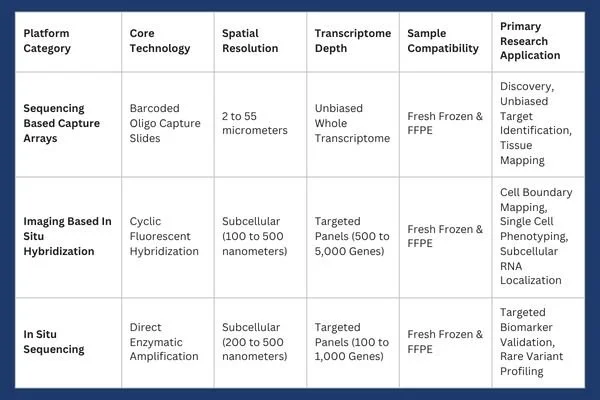
In contrast, imaging based platforms achieve subcellular resolution. These systems utilize targeted fluorescent probe panels to detect individual mRNA molecules within defined cell boundaries. 4
Laboratory Adoption Considerations and Technical Protocols
Successfully establishing a spatial transcriptomics workflow within a biomedical laboratory requires addressing sample preservation, assay selection, and computational data processing infrastructure.
Sample Preparation: FFPE vs Fresh Frozen Tissue
Tissue sample preparation remains a critical variable influencing spatial assay performance.
Fresh Frozen (FF) Tissue: Preserves high-quality RNA integrity, making it optimal for whole transcriptome sequencing-based profiling. However, collecting and preserving fresh frozen samples requires continuous cold-chain storage, which can be difficult to maintain across multisite clinical trial networks.
Formalin Fixed Paraffin Embedded (FFPE) Tissue: Represents the standard preservation format for historical clinical pathology archives. While formalin fixation induces crosslinking and RNA degradation over time, modern probe-based capture chemistries allow robust RNA transcript detection directly from archival FFPE tissue blocks.
Resolution and Multiplexing Tradeoffs
Laboratories must balance transcriptomic breadth against spatial resolution based on specific experimental requirements.
Whole Transcriptome Array Profiling: Measures overall gene expression across thousands of mRNA species across array capture spots. While comprehensive, spatial resolution on standard arrays may encompass multiple adjacent cells per spot, requiring bioinformatic deconvolution to estimate individual cell contributions.
Subcellular Single-Molecule Imaging: Uses targeted, multiplexed probes to achieve resolution down to the hundreds of nanometers. These methods allow direct visualization of intracellular transcript localization within specific organelles or near cell membranes, but require preselecting specific gene-target panels.
Computational Pipelines and Data Scale
Spatial transcriptomics experiments produce extensive datasets combining high-resolution multi-channel image stacks with dense molecular expression matrices. Processing these datasets requires specialized bioinformatic tools capable of handling image registration, cell boundary segmentation, background signal suppression, and spatial neighborhood clustering.
Market Trajectory and Commercial Dynamics
Driven by demand for targeted therapies and precision oncology, the spatial biology market is growing rapidly. Commercial market reports project strong expansion across reagents, instruments, and analytical software through the coming decade.
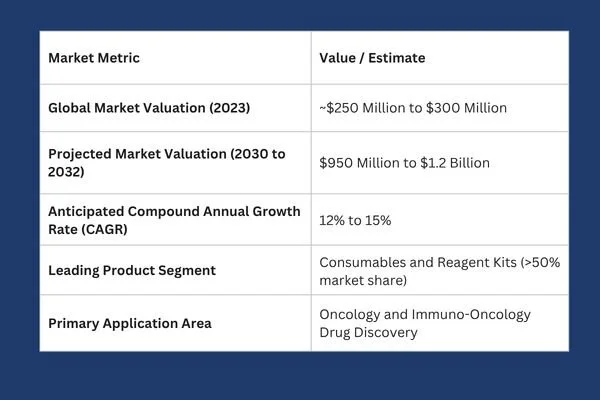
Key drivers powering this commercial expansion include automated tissue-processing instruments, decreasing sequencing costs, improved single-cell segmentation algorithms, and expanding spatial foundation models for automated pathology analysis.
Future Directions: Multimodal Spatial Omics and Artificial Intelligence
The field of spatial biology is rapidly moving beyond single-analyte transcript mapping toward integrated multimodal measurements. Modern experimental platforms enable concurrent spatial profiling of transcripts, functional proteins, epigenetic chromatin accessibility, and small-molecule metabolites within the exact same tissue section.
Concurrently, artificial intelligence models and spatial foundation frameworks are reshaping tissue analytics. Advanced machine learning algorithms can now correlate standard hematoxylin and eosin (H&E) pathology images with underlying spatial gene expression patterns. By training neural networks on matched spatial transcriptomic datasets, researchers can predict localized molecular signatures directly from routine histological slides, unlocking deep biological insight from archival clinical pathology banks.
As these technologies continue to mature and protocol costs decline, spatially resolved biology will evolve from an exploratory research tool into a mandatory standard across pharmaceutical target validation, safety testing, and translational medicine. By preserving the architectural landscape of human tissue, spatial omics is establishing a more accurate foundation for precision drug development.
1 National Library of Medicine, Zhai, Chen & Deng. Researchers can view both the identity of the cell and its exact cellular neighborhood, linking transcriptomic expression directly to tissue histology. pmc.ncbi.nlm.nig.gov
2 Illumina, "What is transcriptomics? A complete guide to gene expression analysis, July 29, 2026. Comprehensive guides on transcriptomic workflows can be found via Illumina transcriptomics overview. illumina.com
3 Atlantis Bioscience, Discovering Solutions for Translational Science. Researchers seeking technical validation tools, reagents, and spatial profiling solutions can explore resources hosted at Atlantis Bioscience. atlantisbioscience.com
4 University of Wisconsin-Madison, Biotechnology Center Gene Expression Core. 10xGenomics Xenium in Situ Spatial Platform. Specialized commercial platforms in this space include the 10x Genomics Xenium in situ platform. biotech.wisc.edu S and the Vizgen MERSCOPE platform. vizgen.com
Our expert advisors accelerate your therapeutic candidates from target validation to clinical triumph. Contact Metis Consulting Services today to integrate cutting-edge spatial transcriptomics and precision workflow strategies into your drug discovery pipeline.

What Acceleration of Development Looks Like in Cell and Gene Therapy
Cell and gene therapy represents some of the most transformative areas of modern medicine. These new and exciting innovations aim to repair, replace, or modify genetic material and cells to treat disease at its root cause.

For Metis Consulting Services
By Michael Bronfman
September 1, 2025
Cell and gene therapy represent some of the most transformative areas of modern medicine. These new and exciting innovations aim to repair, replace, or modify genetic material and cells to treat disease at its root cause. For decades, many conditions, such as certain genetic disorders, advanced cancers, or degenerative diseases, offered few options beyond supportive care. Today, cell and gene therapies are offering new hope. What makes this progress even more remarkable is the speed of development. The acceleration of discovery, design, testing, and approval is unlike any other period in pharmaceutical history.
This article examines how acceleration of development is taking place in cell and gene therapy, why it matters, and what challenges remain.
The Foundation of Acceleration
Progress and practical necessity are forming the foundation of accelerated development in both scientific and. Molecular biology, sequencing technologies, and cell culture methods have all experienced significant advances, reducing the time required to identify disease targets and design therapies. At the same time, patients with life-threatening illnesses cannot afford to wait decades for new treatments. This combination of scientific readiness and urgent medical need has prompted researchers, companies, and regulators to accelerate their efforts.
The path from concept to treatment has traditionally been a slow process. Historically, developing a new pharmaceutical product could take ten to fifteen years. Much of this time was spent in early research, animal testing, and multiple stages of clinical trials. In cell and gene therapy, this timeline has been shortened through better tools, closer collaboration, and more flexible regulatory pathways.
Scientific Tools Driving Speed
One of the most important drivers of acceleration is the set of scientific tools available to researchers. Whole genome sequencing now allows scientists to identify mutations linked to disease in a matter of weeks rather than years. The cost of sequencing has fallen dramatically, which makes it possible to analyze large patient populations and pinpoint the genetic roots of rare conditions.
Gene editing tools such as CRISPR-Cas9 have also transformed development. Before these tools, altering genetic material was complicated, imprecise, and slow. Now, researchers can design edits with remarkable accuracy. This makes it faster to create cell models, test therapeutic approaches, and move into preclinical development.
Advanced cell culture technologies and bioreactors also play a role. Manufacturing therapeutic cells once required painstaking manual methods. Today, automated and scalable systems allow for faster and more consistent production of cells that can be used in therapies.
Collaboration Across Sectors
Acceleration is not only a matter of scientific tools. It also depends on collaboration across sectors. Cell and gene therapy requires expertise from biology, chemistry, engineering, medicine, and regulatory science. Companies have learned that they cannot work in isolation. Instead, partnerships between academic institutions, biotechnology firms, large pharmaceutical companies, and government agencies are essential.
Academic groups often generate the first ideas and proof-of-concept studies. Biotechnology firms then specialize in translating these discoveries into viable products. Large pharmaceutical companies provide resources for clinical trials, manufacturing, and distribution. Regulatory agencies create frameworks that balance safety with speed. Together, these players create an environment in which therapies can progress more quickly than in the past.
Public-private partnerships have been especially valuable. By sharing data, patient registries, and trial results, the entire community reduces duplication of effort and builds collective knowledge. This culture of openness is critical in a field where patient numbers are often small and every dataset matters.
Regulatory Innovation
Another factor in acceleration is regulatory innovation. Traditional drug development was designed for medicines that would be used by millions of patients. Cell and gene therapies are often targeted to rare diseases with small patient populations. Regulators have recognized that the standard framework may not apply in these cases.
In the United States, the Food and Drug Administration has created programs such as the Regenerative Medicine Advanced Therapy Designation. This allows promising therapies to receive closer guidance, rolling review of data, and potential priority review. In Europe, the European Medicines Agency has similar pathways, including the PRIME scheme. These approaches do not remove the requirement for safety and efficacy, but they allow data to be submitted in stages and evaluated more quickly.
Post-marketing commitments are also an important part of the regulatory landscape. Because long-term data may not be available at the time of approval, regulators often allow conditional approval while requiring companies to continue gathering information. This model supports faster access for patients while ensuring continued oversight.
Manufacturing as a Driver of Speed
Manufacturing has traditionally been one of the slowest steps in cell and gene therapy. Producing living cells or viral vectors at clinical and commercial scale is complex. In the early years, many therapies were made almost by hand, which limited capacity and slowed development.
Recent advances are changing this picture. Closed system bioreactors reduce contamination risk and allow for automated production. Improvements in viral vector production yield higher amounts of material in shorter times. Standardization of processes across laboratories and companies also helps reduce delays.
In addition, digital technologies are being used to monitor and optimize manufacturing in real time. This allows rapid adjustments and reduces the likelihood of production failures. Faster and more reliable manufacturing means that clinical trials can begin sooner and scale up more quickly if results are positive.
Patient Engagement
Patients are not simply recipients of cell and gene therapies. They are active participants in the acceleration process. For many rare diseases, patients and advocacy groups are often the driving force behind research funding, trial recruitment, and awareness campaigns.
Engaged patients help researchers design better trials. They provide insight into meaningful outcomes beyond laboratory measurements. They also assist with identifying trial participants, which is especially important when the patient population is very small.
This level of involvement speeds up both the research and the clinical stages. It also ensures that the therapies being developed address real patient needs.
The Role of Data
The acceleration of development is closely tied to the power of data. Electronic health records, genomic databases, and real-world evidence create opportunities to study diseases and track treatment outcomes more efficiently.
Big data approaches allow researchers to identify patterns and predict which patients are most likely to benefit from a given therapy. Adaptive trial designs use data in real time to adjust study parameters, which shortens timelines and increases efficiency.
Data sharing across organizations is also critical. Rare disease research often involves small numbers of patients scattered around the world. Pooling this information accelerates the learning process and reduces duplication of effort.
Case Examples of Accelerated Therapies
Several therapies highlight how accelerated development is reshaping medicine.
Chimeric antigen receptor T cell therapy, known as CAR T therapy, moved from early clinical trials to approval in just a few years. These therapies involve collecting a patient's own T cells, engineering them to attack cancer cells, and reinfusing them. The speed of development was driven by strong clinical results, regulatory flexibility, and urgent patient need.
Another example is gene therapy for spinal muscular atrophy. Once considered untreatable, this disease now has an approved therapy that delivers a functional copy of the defective gene. Development was accelerated by breakthroughs in vector technology, rapid trial enrollment, and close collaboration with regulators.
These examples show that acceleration is not only possible but already happening.
Challenges That Remain
Despite the progress, challenges remain. Safety is always the highest concern. Editing genes or modifying cells carries risks, and long-term monitoring is essential. While acceleration is important, it cannot come at the cost of patient well-being.
Manufacturing and supply chain issues continue to be difficult. Even with improved systems, scaling up to meet demand is not easy. The cost of therapies remains very high, which raises questions about access and sustainability.
Global harmonization is still lacking, even though regulatory pathways are evolving. A therapy could be approved in one region or country and still face delays in others. Greater international coordination could reduce these barriers.
Finally, ethical questions around gene editing and long-term effects require ongoing discussion. Acceleration must be balanced with careful consideration of broader implications.
The Future of Accelerated Development
Looking forward, acceleration of development in cell and gene therapy is likely to continue. New tools such as base editing and prime editing promise even greater precision. Advances in stem cell biology may expand the range of treatable diseases. Manufacturing will become more standardized and less costly.
Regulatory agencies are learning from early experiences and may refine their frameworks to support even faster yet responsible approvals. Global collaboration will likely grow as patient groups, researchers, and companies work across borders.
Ultimately, the greatest measure of acceleration will be patient outcomes. The goal is not speed for its own sake, but faster relief for individuals and families facing devastating conditions. When development accelerates responsibly, it translates into more lives improved and more diseases transformed from untreatable to manageable.
Acceleration of development in cell and gene therapy is one of the most significant trends in modern medicine. The quick progress faster than ever before, is being driven by scientific advances, collaboration, regulatory innovations, improved manufacturing, patient engagement, and the power of data. While challenges remain, the direction is clear. Diseases once thought incurable are now within reach of treatment. The future of medicine will be defined not only by what we can achieve, but also by how quickly we can achieve it responsibly.
The acceleration of development is not simply about speed. It is about building a system that can respond to urgent patient needs, integrate scientific discovery, and deliver safe and effective therapies in record time. Cell and gene therapy demonstrate that this vision is possible and that the future of healthcare is being reshaped today.
As the landscape continues to evolve, staying ahead requires deep expertise and strategic insight. At Metis Consulting Services, we specialize in helping organizations navigate these complexities and harness the full potential of this groundbreaking field.
To learn more about our services and how we can partner with you, please see our website: metisconsultingservices.com or email us at hello@metisconsultingservices.com.