
In Vivo CAR-T Cell Therapy: Rewriting the Rules of Cancer Treatment
This week in the Guardrail, we explore how in vivo CAR-T cell therapy is shifting the paradigm of genetic medicine by reprogramming a patient’s immune system directly inside their body.
By Michael Bronfman
The way we treat cancer is changing fast, and one of the biggest stories in medicine right now is happening inside the human body rather than inside a lab.
For years, doctors have used a groundbreaking treatment called CAR-T cell therapy to fight stubborn blood cancers. It works by taking a patient's own immune cells, giving them a genetic upgrade, and putting them back into the body to hunt down cancer cells. While this therapy has saved many lives, it is extremely hard to make, takes weeks to process, and costs a fortune.
Now, scientists are testing a new approach called in vivo CAR-T cell therapy. Instead of taking cells out of the body to upgrade them, this new method delivers the genetic instructions directly into the patient with a simple injection. It turns the patient's own body into the laboratory.
If this technology succeeds in human trials, it could make life-saving cancer treatments faster, cheaper, and available to millions of people around the world.
What Is Traditional CAR-T Cell Therapy?
To understand why this new discovery is such a big deal, it helps to look at how traditional CAR-T therapy works.
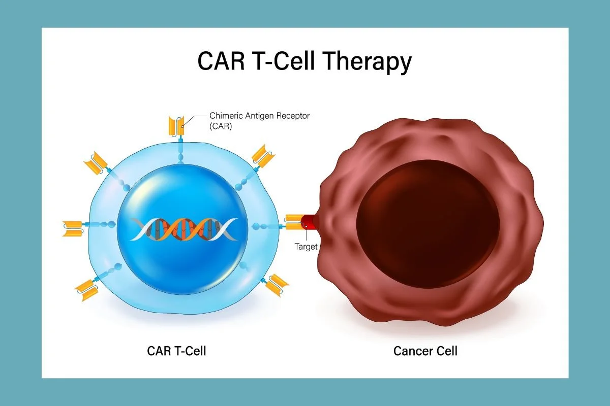
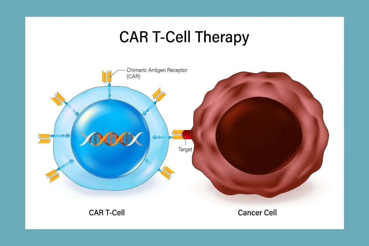
CAR stands for Chimeric Antigen Receptor. T-cells are a type of white blood cell in your immune system. You can think of T-cells as the body's internal security guards. They patrol your bloodstream, looking for infected cells or abnormal cells, and destroy them before they can cause harm.
However, cancer cells are tricky. They often disguise themselves so the immune system cannot see them. To overcome this, scientists figured out a way to retrain T-cells.
Here is the traditional step-by-step process:
Blood Collection: Doctors hook the patient up to a special machine to remove their blood, separate out the T-cells, and return the rest of the blood.
Shipping to a Specialized Lab: The harvested T-cells are frozen and shipped to a high-tech facility known as a Good Manufacturing Practice facility.
Genetic Engineering: In the lab, scientists use a harmless virus to insert a new gene into the T-cells. This gene tells the cells to grow special hooks on their surface called Chimeric Antigen Receptors. These hooks allow the T-cells to lock onto a specific protein found on cancer cells.
Cell Multiplication: The lab grows millions of these upgraded CAR-T cells over several weeks.
Chemotherapy: Before receiving the new cells, the patient goes through chemotherapy to clear out some of their old immune cells and make room for the upgraded ones.
Reinfusion: The new CAR-T cells are shipped back to the hospital and infused into the patient's bloodstream.
Once inside, these engineered T-cells act like guided missiles. They seek out cancer cells, lock onto them with their new receptor hooks, and destroy them.
The Big Problems with Traditional CAR-T
While traditional CAR-T therapy has cleared cancer in patients who had no other options left, the current system has massive drawbacks.
1. Time Limits
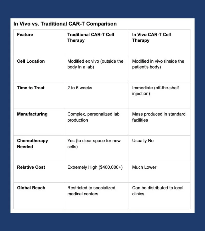
Making CAR-T cells outside the body takes anywhere from two to six weeks. For someone with fast-growing leukemia or lymphoma, waiting weeks for a treatment can be dangerous or even fatal.
2. High Costs
Because every batch of CAR-T cells must be custom-made for one specific individual in a sterile lab, the cost is enormous. A single treatment can cost between four hundred thousand and five hundred thousand dollars, not including hospital stays and extra medical care.
3. Complex Logistics
Shipping living human cells across the country under frozen conditions requires advanced temperature-controlled transport. If anything goes wrong during shipping or manufacturing, the batch can be ruined, forcing the patient to start over.
4. Limited Access
Because the process requires specialized hospitals and advanced manufacturing labs, CAR-T therapy is mostly available in wealthy countries and large academic medical centers. Most people in developing nations or rural areas cannot get access to it.
The In Vivo Breakthrough: How It Works
This is where in vivo CAR-T cell therapy comes in. The phrase in vivo means inside the living body. Instead of taking cells out, modifying them in a lab, and putting them back, in vivo therapy delivers the genetic instructions directly into the patient using a single injection.
Scientists do this with special delivery vehicles called nanoparticles or modified viruses.
Here is how the in vivo process works:
Building the Delivery Vehicle: Scientists create tiny bubbles made of fats, known as lipid nanoparticles, or use modified viral shells. Inside these tiny bubbles, they pack genetic instructions written in mRNA or DNA.
Adding the Navigation System: Scientists attach special targeting molecules to the outside of the nanoparticle. These molecules act like a GPS, ensuring the nanoparticle only attaches to T-cells and ignores other cells like liver or lung cells.
Direct Injection: The patient receives a simple IV drip or injection containing these targeted nanoparticles.
Reprogramming Inside the Body: The nanoparticles travel through the bloodstream, attach to the patient's T-cells, and deliver the genetic instructions.
Cancer Hunting: The T-cells read the instructions, start building chimeric antigen receptors on their surface, and immediately begin hunting down cancer cells inside the body.
This simple shift removes the need for cell extraction, lab manufacturing, complex freezing shipping lines, and heavy chemotherapy prep.
What the Research Shows
Recent preclinical studies have shown promising results using in vivo cell engineering.
Research in animal models demonstrates that mRNA delivered by lipid nanoparticles can successfully target T-cells inside living mice. Within days of injection, the mice generated functional CAR-T cells that successfully targeted and shrank tumors.
Researchers are also exploring precise gene editing tools like CRISPR inside the body. Instead of just adding a new gene temporarily, in vivo gene editing can permanently alter the T-cells or remove genes that cause cellular exhaustion, allowing the immune cells to fight cancer longer.
Scientists are publishing new findings regularly on research platforms like Atlantis Bioscience and medical news outlets like STAT News. These studies highlight how in vivo engineering could cut production times from weeks down to zero, turning a custom surgical procedure into an off the shelf pharmacy medication.
Potential Benefits Beyond Cancer
While cancer treatment is the main focus, in vivo CAR-T cell therapy could treat many other diseases.
Autoimmune Diseases: In conditions like lupus or multiple sclerosis, the immune system mistakenly attacks healthy tissue. Scientists are testing CAR-T cells designed to clear out malfunctioning immune cells, effectively resetting the immune system.
Heart Disease: Researchers have used in vivo CAR-T cells in animal models to target and remove scar tissue in damaged hearts, helping restore heart function after a heart attack.
Infectious Diseases: Modified immune cells could be trained to clear out persistent viral infections like HIV that hide inside human tissues.
Organ Transplants: Engineered regulatory immune cells could prevent organ rejection without requiring lifelong immune-suppressing drugs.
Challenges Ahead
While in vivo CAR-T therapy is exciting, scientists still need to solve several technical challenges before it becomes widely available for human patients.
1. Off-Target Effects
The delivery vehicle must be extremely accurate. If a nanoparticle delivers its genetic payload to the wrong cell type, like liver cells or brain cells, it could cause unintended side effects.
2. Overactive Immune Reaction
When CAR-T cells attack cancer cells, they release signal proteins called cytokines. If too many CAR-T cells activate at once inside the body, it can cause a dangerous condition called cytokine release syndrome, which causes high fevers and dangerous drops in blood pressure. Controlling the speed and strength of in vivo reprogramming is vital for safety.
3. Immune Rejection of Delivery Vehicles
The human immune system might spot the nanoparticles or viral vectors and destroy them before they reach the T-cells. Finding materials that can sneak past the body's defenses without causing an allergic reaction requires careful design.
4. Dosing and Control
With traditional CAR-T, doctors know exactly how many modified cells they put back into the patient. With in vivo therapy, it is harder to predict how many T-cells will pick up the gene instructions and how long those instructions will last.
The Road Ahead
In vivo CAR-T cell therapy represents a major shift in modern medicine. By turning the human body into its own medicine factory, scientists are opening the door to a future where genetic treatments are no longer rare luxury procedures reserved for a few, but standard therapies available at local healthcare clinics.
Clinical trials in humans are beginning to take off, testing different delivery vehicles and gene editing tools. Over the next decade, as researchers refine these methods, in vivo cell engineering could fundamentally reshape how we treat cancer, autoimmune conditions, and chronic heart disease.
With Metis Consulting Services you streamline your strategic roadmap, overcome complex regulatory hurdles, and maximize operational efficiency. Contact Metis Consulting Services today

Direct Cellular Engineering
Direct cellular engineering

This week in the Guardrail, we explore how shifting cellular engineering directly inside the human body could eliminate costly manufacturing delays and revolutionize patient access to life-saving treatments. Are you ready to overcome regulatory and delivery hurdles and bring next-generation cell therapies to market faster? Read on.
By Michael Bronfman
August 24, 2026
Traditional cell therapies have achieved extraordinary accomplishments in modern oncology. Chimeric antigen receptor T cell treatments have altered the prognosis for patients suffering from aggressive hematological malignancies like relapsed leukemias, lymphomas, and multiple myeloma. Despite these clinical achievements, the logistical structure of modern cell therapy remains fundamentally constrained.
The current standard of care relies entirely on an autologous ex vivo manufacturing paradigm. Clinicians must harvest T cells from a patient through leukapheresis, transport those living cells to dedicated Good Manufacturing Practice facilities, genetically reprogram them using viral vectors, expand them over several weeks, and deliver them back to the clinical site for reinfusion.
Manufacturing schedules routinely span three to five weeks. This process presents severe bottlenecks. Throughout this waiting window, rapidly progressing disease can cause patient deterioration or mortality before the engineered cells arrive. Intensive labor, stringent quality control, and specialized infrastructure drive treatment prices to hundreds of thousands of dollars per patient, excluding ancillary medical costs for lymphodepleting chemotherapy and prolonged hospital stays.
The Ex Vivo Bottleneck vs The In Vivo Solution
To understand the revolutionary potential of in situ reprogramming, one must examine the structural restrictions inherent to ex vivo cell manufacturing.
Traditional Ex Vivo Pathway
Patient leukapheresis harvesting
Transport to a specialized GMP lab
Viral engineering and gene insertion
Multi-week ex vivo cellular expansion
Transport back to clinic and reinfusion following patient lymphodepletion
In Vivo Pathway
Direct intravenous infusion of targeted vector formulation
Immediate in situ T cell editing within seconds to hours inside the patient's body
Key Differences in Delivery Logistics
Cell Processing Requirements: Ex vivo protocols demand complex cell isolation, activation, viral transduction, and extensive expansion steps outside the body. In vivo protocols utilize an off-the-shelf synthetic formulation or targeted vector that requires no patient-specific cell manipulation.
Therapeutic Turnaround: Traditional manufacturing requires weeks of processing time, during which patients often require bridging therapies to manage disease burden. In vivo vectors can be reconstituted and administered on the same day the treatment decision is made.
Scale and Health Facilities: Current cell therapies are largely restricted to major academic medical centers that manage cell handling, logistics chain, and complex toxicities. In vivo formulations behave like traditional biologic drugs, opening up administration to regional health centers and community hospitals.
Cellular Fitness and Stemness: When T cells are harvested, stimulated, and cultured ex vivo for extended durations, they frequently undergo metabolic stress and premature exhaustion. Cells engineered directly inside the living organism retain their natural physiological state, naive phenotypes, and proliferative potential.
Technical Delivery Platforms
Directing genetic material to specific immune cell subsets within a complex systemic environment requires sophisticated delivery systems that can navigate biological barriers without triggering unwanted off-target effects.
Targeted Lipid Nanoparticles
Lipid nanoparticles, similar to those validated during global mRNA vaccine deployment, serve as a non-viral platform for in vivo reprogramming. Standard LNPs naturally accumulate in the liver due to apolipoprotein E adsorption. To redirect these particles specifically toward T lymphocytes, researchers modify the outer lipid shell with targeted ligands, such as monoclonal antibody fragments or single-chain variable fragments that bind surface receptors like CD3, CD4, or CD5.
The structural makeup consists of:
Outer Shell: Targeted ligands (anti-CD3 or anti-CD5 antibody fragments) and PEGylated lipids for target specificity and stability.
Internal Core: Ionizable lipids and structural phospholipids surrounding the encapsulated payload.
Payload: Messenger RNA encoding the CAR construct or CRISPR-Cas editing machinery.
Once administered intravenously, these surface-modified LNPs recognize circulating and tissue-resident T cells and induce receptor-mediated endocytosis. Inside the cell, the ionizable lipids respond to the acidic endosomal environment, damage the membrane, and release the encapsulated messenger RNA into the cytoplasm.
The cytoplasm then translates the mRNA payload into CAR proteins that translocate to the cell membrane. Because mRNA does not integrate into the genomic DNA, expression is transient, lasting from several days to a few weeks. This time-based control offers a major safety advantage, allowing clinicians to titrate dosage and avoid permanently immune-altered states.
Engineered Viral Vectors
For applications requiring permanent genetic integration, researchers are actively refining pseudotyped viral delivery systems. Standard viral vectors, such as adeno-associated viruses or lentiviruses, exhibit broad tropism and infect various cell types indiscriminately.
To overcome this, engineers mutate the native viral envelope proteins to abolish their natural binding capabilities. They then engineer the viral surface to display targeting domains specific to T cell surface markers.
Receptor-targeted lentiviral vectors can selectively transduce CD8-positive cytotoxic lymphocytes or CD4-positive helper lymphocytes directly within lymph nodes and the spleen. This permanent integration guarantees that as the reprogrammed T cells divide in response to tumor antigens, their daughter cells inherit the CAR gene, providing long-term immunological memory.
In Vivo Site-Specific Genomic Editing
A major milestone in cellular engineering is securing precise gene insertion in vivo without random viral integration. Recent developments use dual-vector platforms that combine enveloped delivery vehicles with adeno-associated viruses to execute site-specific knock-ins directly inside endogenous T cells.
The site-specific knock-in mechanism proceeds through distinct biological phases:
Targeting: Guide RNA directs the Cas9 nuclease to a designated cut site within Exon 1 of the TRAC locus.
Cleavage: Double-strand DNA breaks are introduced precisely at the target site, knocking out the endogenous T cell receptor.
Integration: Homology-directed repair templates insert the synthetic CAR gene sequence into the double-strand break point.
Expression: The integrated CAR gene is transcribed directly under the control of native promoter elements.
Furthermore, conducting this site-specific knockout and knock-in process entirely in vivo preserves T cell stemness. Recent preclinical findings show that T cells engineered inside living tissue express higher levels of stem cell memory markers versus traditional lab-engineered cell products, yielding superior persistence and deeper tumor-clearing capabilities in both hematological and solid tumor models.
Primary Applications and Preclinical Findings
Direct immune cell editing is expanding into wider medical disciplines. Oncology remains the primary target for in vivo CAR technologies.
Hematological Cancers
Primary Target Indications: Leukemias, Lymphomas, Multiple Myeloma
Biological Mechanism: Transient or permanent targeting of CD19 or CD20 on malignant B cell populations
Solid Tumors
Primary Target Indications: Epithelial carcinomas, Glioblastomas
Biological Mechanism: Multi-antigen targeting combined with microenvironment remodeling factors
Autoimmune Diseases
Primary Target Indications: Lupus, Rheumatoid Arthritis
Biological Mechanism: Transient depletion of autoreactive B cells or generation of regulatory T cells
Tissue Fibrosis
Primary Target Indications: Cardiac scarring, Liver cirrhosis
Biological Mechanism: Anti-FAP CARs targeting activated fibroblasts to reverse tissue scarring
Hematological and Solid Tumors
In preclinical models, targeted LNPs delivering anti-CD19 CAR mRNA have rapidly generated functional CAR T populations in circulating blood and lymphatic tissue. These in situ generated cells cleared systemic tumor burdens with efficiency matching or exceeding ex vivo controls.
Dual vector gene insertion strategies target the TRAC locus and achieve therapeutic levels of CAR T cells in vivo, successfully clearing aggressive leukemias and multiple myeloma in humanized animal models with a single systemic injection.
Autoimmune and Fibrotic Diseases
The transient nature of non-viral mRNA delivery makes in vivo CAR generation uniquely suited for non-oncology indications where permanent cell modification is unnecessary or hazardous.
Cardiac and Tissue Fibrosis: Pathological fibrosis in heart disease and liver conditions is driven by overactive, activated fibroblasts expressing Fibroblast Activation Protein. Research using CD5-targeted LNPs carrying anti-FAP CAR mRNA showed that a single systemic infusion could reprogram T cells to selectively eliminate activated fibroblasts, significantly reducing cardiac and hepatic fibrosis and restoring organ function.
Autoimmune Disorders: In severe autoimmune conditions like systemic lupus erythematosus, transient in vivo depletion of the pathogenic B cell population resets the immune system. Once the transient CAR expression dissipates, naive B cells repopulate the compartment without causing permanent humoral immunodeficiency.
Off-Target Transduction and Tissue Tropism
A central safety requirement for overall vector administration is absolute target specificity. If a viral or nanoparticle vector encapsulates or transduces off-target tissues like hepatocytes, endothelial cells, or healthy parenchymal tissue, the host cells may express chimeric antigen receptors on their surface or suffer unintended genetic editing.
To reduce off-target risks, developers use dual-targeting strategies. This includes combining surface antibody functionalization with cell type-specific promoter sequences, making sure that even if a vector enters a non-immune cell, the synthetic genetic payload remains transcriptionally silent.
Academic References
For additional information about the core mechanisms and translational data of in vivo cellular engineering, refer to the following publications:
Volta, L. (2026). In vivo generation of CAR T cells: biology, delivery platforms, clinical promise, and translational challenges. Blood Immunology & Cellular Therapy, 2(1), 100027.
Nyberg, W. A., Bernard, P. L., Ngo, W., et al. (2026). In vivo site-specific engineering to reprogram T cells. Nature, 639, 10235.
Rurik, J. G., Tombácz, I., Yadegari, A., et al. (2022). CAR T cells produced in vivo to treat cardiac injury. Science, 375(6578), 91-96.
Klichinsky, M., Ruella, M., Shestova, O., et al. (2020). Human chimeric antigen receptor macrophages for cancer immunotherapy. Nature Biotechnology, 38(8), 947-953.
To learn more about cellular processing standards and bio manufacturing logistics, explore technical guidelines supplied by the International Society for Cell & Gene Therapy and regulatory insights at the U.S. Food and Drug Administration Cellular & Gene Therapy Products Portal.
Spatial Omics and Single-Cell Biology: Mapping the Next Generation of Precision Therapeutics
Today, we examine how spatial omics and single-cell biology are overcoming the limitations of bulk sequencing by mapping gene activity directly within intact tissue architecture. Discover how this architectural shift is accelerating target discovery, refining biomarker validation, and powering the next generation of precision therapeutics.
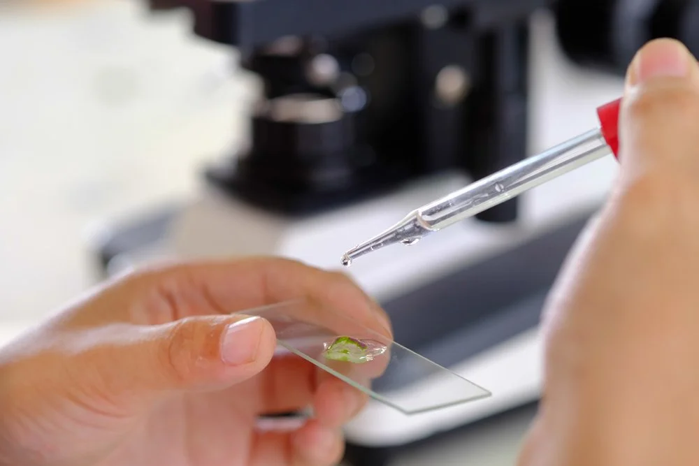
This week in the Guardrail… we examine how spatial omics and single-cell biology are overcoming the limitations of bulk sequencing by mapping gene activity directly within intact tissue architecture. Discover how this architectural shift is accelerating target discovery, refining biomarker validation, and powering the next generation of precision therapeutics.
By Michael Bronfman
August 17, 2026
Traditional genomic and transcriptomic analyses have long served as the foundation of biomedical research, yet they carry a fundamental structural limitation. Standard sequencing protocols require researchers to grind up heterogeneous tissue samples to extract their DNA or RNA. While this bulk extraction yields valuable genetic readouts, it completely destroys the spatial architecture of the tissue sample. The homogenization process blends thousands or millions of distinct cells into a single average measurement. This leaves researchers unable to observe how individual cell types were originally organized, how they communicated through local signaling, or how they interacted within their microenvironment.
Spatial transcriptomics and single-cell biology directly overcome this limitation. By enabling scientists to profile gene activity directly within an intact tissue section, these combined methodologies preserve critical spatial coordinates alongside comprehensive molecular signatures. By bridging single-cell sequencing with high-resolution optical imaging, spatial omics maps the exact location of individual cell populations while detailing their corresponding gene expression profiles inside native tissue architecture.
This technical shift is accelerating preclinical research, refining target discovery, and transforming drug development strategies across major therapeutic areas.
The Technological Shift: Moving Beyond Bulk Averages
For decades, drug discovery teams relied heavily on bulk sequencing data to identify therapeutic targets. Bulk measurements, however, routinely mask low-abundance cell populations or localized transcriptional shifts. A candidate drug target that appears minimally expressed across an entire tumor lysate may actually be highly expressed within a localized cluster of aggressive cells at the invasive tumor margin.
Single-cell RNA sequencing introduced higher resolution by isolating individual cells prior to library preparation, enabling researchers to classify distinct cell subtypes and rare phenotypes. Yet, the physical dissociation step required for single-cell RNA sequencing severs extracellular matrix contacts and destroys spatial organization.
Spatial omics platforms bridge this remaining gap. By utilizing spatially barcoded solid-phase capture arrays or multiplexed in situ hybridization, these technologies capture mRNA transcripts directly from intact tissue sections. 1
Transformative Impact Across Key Therapeutic Areas
Integrating spatial profiling into preclinical drug development pipelines provides unprecedented clarity into complex human diseases, directly improving target validation and risk assessment.
Oncology and the Tumor Microenvironment
Cancerous tissue is exceptionally complex, consisting of malignant subclones, infiltrating immune cells, stromal fibroblasts, blood vessels, and dense extracellular matrix components. Spatial omics allows oncology researchers to deconstruct the tumor microenvironment with high spatial resolution.
Immune Exclusion Zones: Spatial profiling explains why checkpoint inhibitors succeed in certain regions of a tumor while failing in others. Researchers can identify physically restricted immune cells trapped within dense fibrotic borders, which prevent them from penetrating the core tumor tissue.
Therapeutic Resistance Niches: Tumor regions located near hypovascularized or hypoxic cores display distinct stress-response signatures. Mapping these microdomains clarifies how localized environments shield malignant subclones from systemically administered therapies.
Tertiary Lymphoid Structures: Identifying the presence, cellular composition, and spatial layout of tertiary lymphoid structures within solid tumors offers strong predictive value for immunotherapy response. This enables clinical teams to stratify patient cohorts more effectively during early clinical trials.
Neuroscience and Structural Brain Mapping
The central nervous system depends entirely on an organized spatial architecture, in which local neuronal circuits govern functional signals. Bulk sequencing of brain tissue offers limited actionable insight into circuit-level pathologies.
Neuronal Circuit Profiling: Spatial transcriptomics maps the complex laminar organization of the cerebral cortex, charting gene signatures across distinct cortical layers and subcortical structures.
Neurodegenerative Pathology Progression: In conditions such as Alzheimer disease or Parkinson disease, pathological features such as amyloid plaques and neurofibrillary tangles form in localized regions. Spatial profiling allows researchers to evaluate transcriptomic shifts occurring in microglial cells and neurons immediately adjacent to these protein aggregates, comparing them directly to distant healthy tissue.
Blood-Brain Barrier Integrity: Mapping the vascular parenchymal interface helps researchers evaluate how local inflammatory signals compromise active transport across the blood-brain barrier. This aids the rational design of central nervous system drug delivery vehicles.
Immunology and Inflammatory Pathology
Immune responses depend on coordinated cellular migration and localized paracrine signaling within specialized tissue structures.
Lymph Node Architecture: Spatial methods map how immune cell subsets, including dendritic cells, helper T cells, and cytotoxic lymphocytes, reorganize within follicular and paracortical regions during antigen presentation or autoimmune activation.
Autoimmune Niches: In chronic inflammatory conditions such as rheumatoid arthritis or Crohn disease, spatial omics identifies local cellular niches that drive tissue damage. This uncovers localized cytokine networks that systemic blood profiling fails to detect.
Integrating Spatial Omics into the Drug Discovery Pipeline
Pharmaceutical R&D organizations are transitioning from bulk tissue profiling to spatially resolved experimental workflows. This workflow integration enhances multiple stages of early drug development.
Target Identification and Validation
By overlaying high-resolution transcript profiling onto tissue histology, discovery teams can verify whether a target is expressed specifically within disease-driving cells or broadly across healthy tissue. Target evaluation at the tissue microdomain level reduces off-target toxicity risks early in discovery.2
Pharmacodynamics and Tissue Distribution
Assessing candidate drug efficacy requires confirming that a therapeutic agent reaches target cells at effective concentrations within complex tissue. Combining spatial transcriptomics with mass spectrometry imaging or spatial proteomics allows research teams to map drug molecule concentration alongside downstream gene expression changes in the exact same tissue section.
Biomarker Discovery for Clinical Stratification
Phase 2 and Phase 3 clinical trial failures frequently stem from unaddressed patient heterogeneity. Analyzing intact patient biopsy tissue reveals spatial biomarkers, such as cell-to-cell proximity scores or immune infiltration indices, that identify patient subgroups most likely to achieve clinical response.3
Comparative Analysis of Primary Spatial Omics Technologies
Modern spatial biology platforms fall into two main technical categories: sequencing-based spatial capture arrays and imaging-based in situ hybridization technologies. Selecting the appropriate technology depends on research goals, resolution requirements, and target gene panel depth.Sequencing-based capture platforms, such as array-based transcript mapping systems, provide unbiased, high-throughput coverage across the entire genome. These systems are ideal for exploratory target discovery where relevant biological pathways are not yet fully defined.
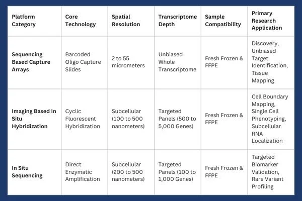
In contrast, imaging based platforms achieve subcellular resolution. These systems utilize targeted fluorescent probe panels to detect individual mRNA molecules within defined cell boundaries. 4
Laboratory Adoption Considerations and Technical Protocols
Successfully establishing a spatial transcriptomics workflow within a biomedical laboratory requires addressing sample preservation, assay selection, and computational data processing infrastructure.
Sample Preparation: FFPE vs Fresh Frozen Tissue
Tissue sample preparation remains a critical variable influencing spatial assay performance.
Fresh Frozen (FF) Tissue: Preserves high-quality RNA integrity, making it optimal for whole transcriptome sequencing-based profiling. However, collecting and preserving fresh frozen samples requires continuous cold-chain storage, which can be difficult to maintain across multisite clinical trial networks.
Formalin Fixed Paraffin Embedded (FFPE) Tissue: Represents the standard preservation format for historical clinical pathology archives. While formalin fixation induces crosslinking and RNA degradation over time, modern probe-based capture chemistries allow robust RNA transcript detection directly from archival FFPE tissue blocks.
Resolution and Multiplexing Tradeoffs
Laboratories must balance transcriptomic breadth against spatial resolution based on specific experimental requirements.
Whole Transcriptome Array Profiling: Measures overall gene expression across thousands of mRNA species across array capture spots. While comprehensive, spatial resolution on standard arrays may encompass multiple adjacent cells per spot, requiring bioinformatic deconvolution to estimate individual cell contributions.
Subcellular Single-Molecule Imaging: Uses targeted, multiplexed probes to achieve resolution down to the hundreds of nanometers. These methods allow direct visualization of intracellular transcript localization within specific organelles or near cell membranes, but require preselecting specific gene-target panels.
Computational Pipelines and Data Scale
Spatial transcriptomics experiments produce extensive datasets combining high-resolution multi-channel image stacks with dense molecular expression matrices. Processing these datasets requires specialized bioinformatic tools capable of handling image registration, cell boundary segmentation, background signal suppression, and spatial neighborhood clustering.
Market Trajectory and Commercial Dynamics
Driven by demand for targeted therapies and precision oncology, the spatial biology market is growing rapidly. Commercial market reports project strong expansion across reagents, instruments, and analytical software through the coming decade.
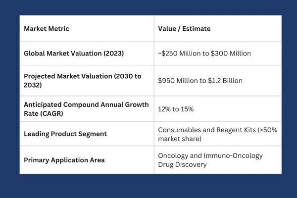
Key drivers powering this commercial expansion include automated tissue-processing instruments, decreasing sequencing costs, improved single-cell segmentation algorithms, and expanding spatial foundation models for automated pathology analysis.
Future Directions: Multimodal Spatial Omics and Artificial Intelligence
The field of spatial biology is rapidly moving beyond single-analyte transcript mapping toward integrated multimodal measurements. Modern experimental platforms enable concurrent spatial profiling of transcripts, functional proteins, epigenetic chromatin accessibility, and small-molecule metabolites within the exact same tissue section.
Concurrently, artificial intelligence models and spatial foundation frameworks are reshaping tissue analytics. Advanced machine learning algorithms can now correlate standard hematoxylin and eosin (H&E) pathology images with underlying spatial gene expression patterns. By training neural networks on matched spatial transcriptomic datasets, researchers can predict localized molecular signatures directly from routine histological slides, unlocking deep biological insight from archival clinical pathology banks.
As these technologies continue to mature and protocol costs decline, spatially resolved biology will evolve from an exploratory research tool into a mandatory standard across pharmaceutical target validation, safety testing, and translational medicine. By preserving the architectural landscape of human tissue, spatial omics is establishing a more accurate foundation for precision drug development.
1 National Library of Medicine, Zhai, Chen & Deng. Researchers can view both the identity of the cell and its exact cellular neighborhood, linking transcriptomic expression directly to tissue histology. pmc.ncbi.nlm.nig.gov
2 Illumina, "What is transcriptomics? A complete guide to gene expression analysis, July 29, 2026. Comprehensive guides on transcriptomic workflows can be found via Illumina transcriptomics overview. illumina.com
3 Atlantis Bioscience, Discovering Solutions for Translational Science. Researchers seeking technical validation tools, reagents, and spatial profiling solutions can explore resources hosted at Atlantis Bioscience. atlantisbioscience.com
4 University of Wisconsin-Madison, Biotechnology Center Gene Expression Core. 10xGenomics Xenium in Situ Spatial Platform. Specialized commercial platforms in this space include the 10x Genomics Xenium in situ platform. biotech.wisc.edu S and the Vizgen MERSCOPE platform. vizgen.com
Our expert advisors accelerate your therapeutic candidates from target validation to clinical triumph. Contact Metis Consulting Services today to integrate cutting-edge spatial transcriptomics and precision workflow strategies into your drug discovery pipeline.
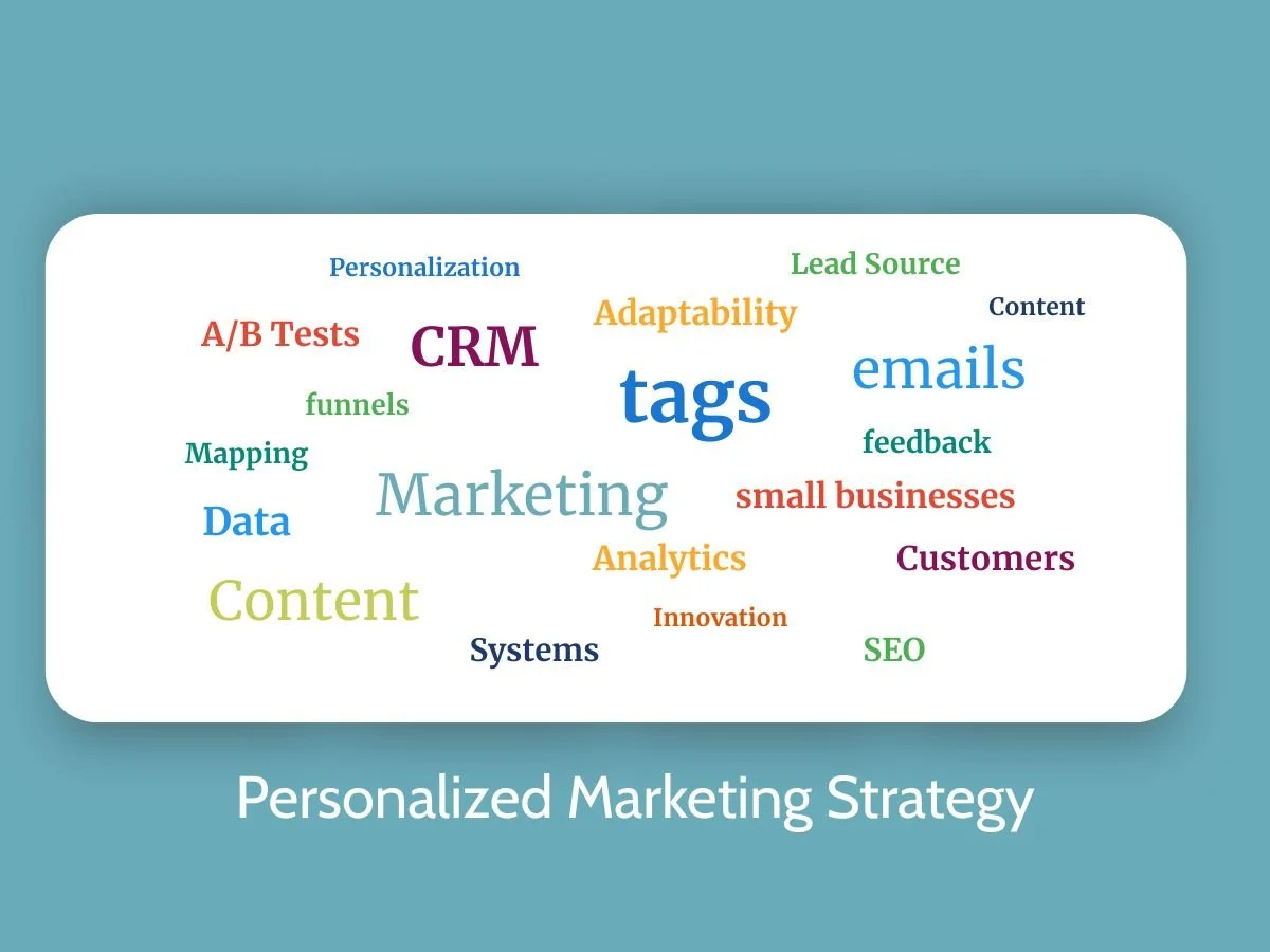
How to Seamlessly Plan and Launch Your Personalized Marketing Strategy
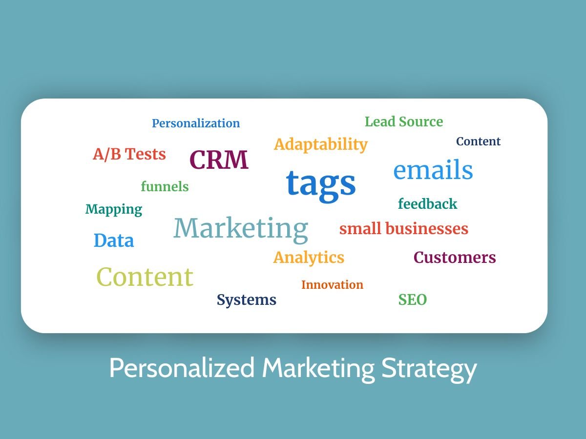
This week, guest Blogger Lance Cody-Valdez discusses a practical framework for small businesses looking to seamlessly plan, test, and scale a content personalization strategy without overwhelming their marketing stack. By taking a phased, data-driven approach, companies can turn complex integrations into a predictable, high-impact engine for growth.
By Lance Cody-Valdez
August 10, 2026
For small business owners, a personalized marketing strategy often sounds simple until content personalization integration starts tugging on every part of the marketing stack. The core challenge is that personalization touches data, tools, and day-to-day workflows at the same time, so quick add-ons can create messy handoffs and inconsistent customer experiences. With a clear project planning approach, personalization becomes a coordinated rollout instead of a patchwork of fixes. The payoff is marketing that feels relevant and stays manageable.
Quick Summary: Planning Content Personalization
● Define clear content personalization goals that align with business outcomes and audience needs.
● Audit your technology stack to confirm data sources, integrations, and delivery capabilities.
● Segment audiences thoughtfully and prioritize data security and privacy from the start.
● Launch in phases with cross-departmental collaboration to reduce risk and improve adoption.
● Track performance metrics consistently to validate impact and guide ongoing optimization.
Where Personalization Fits in Your Marketing Tools
Start with the bigger picture.
Content personalization tools are not a standalone magic button. They sit between the systems that store customer details (your CRM), send messages (email and automation), measure behavior (analytics), and sometimes even trigger offers after a purchase (payments). The core idea is mapping what you already use, then checking what data can actually flow between tools, and which foundational systems matter most for revenue and growth, as outlined in essential small business technology.
This matters because most small businesses have tools, but not a clean system. When only 18 percent of SMBs feel “very confident” in marketing effectiveness, gaps often come from disconnected data and unclear ownership. A simple inventory helps you fix the right problem first.
Think of it like a kitchen setup. You can buy a blender, but you still need ingredients, a power outlet, and a recipe. Your reference list is the checklist: CRM fields, email list source, tracking tags, and purchase history access.
With your stack mapped, you can set goals and confirm compatibility before building personas and pilots.
Build a Simple Personalization Launch Plan
This process helps you turn “we want personalization” into a practical project plan you can actually run. You will define what success looks like, confirm your tools can support it, and launch a small pilot without creating data or ownership headaches.
1. Step 1: Set one clear personalization objective
Start with a single outcome tied to a business goal, like increasing email clicks on a key offer or improving repeat purchases from recent buyers. Define one primary metric and one timeframe so you can tell if personalization worked without debate. This keeps your first rollout focused and easier to resource.
2. Step 2: Confirm tech compatibility and data access
Review which system will be your “source of truth” for customer details, and list the exact fields you need (for example: product category purchased, lead source, last visit date). Then check whether those fields can reliably flow into the channels you will personalize, even if it is a basic import or a simple integration. If the data cannot move cleanly, adjust the objective or simplify the audience rules.
3. Step 3: Build 2 to 3 actionable personas from real signals
Choose segments you can identify with data you already have, such as first-time buyers, high-intent browsers, or loyal repeat customers. For each persona, write: the trigger you will use, the message they should see, and the next action you want them to take. Keep it measurable, since personas are only useful if you can target them consistently.
4. Step 4: Check consent, privacy, and basic data hygiene
Document what data you will use, where it came from, and who can access it, then confirm it aligns with your email and cookie consent practices. Clean up the minimum fields required (naming conventions, missing values, duplicates), so your targeting rules do not misfire. This step prevents “creepy” personalization and reduces broken experiences.
5. Step 5: Run a small pilot and align stakeholders on ownership
Pick one channel and one use case, then run an A/B test or a holdout comparison long enough to see a stable signal. Assign a small working group with clear roles, and select representatives with the ability and willingness to represent stakeholder groups so decisions do not stall across teams. Capture results, feedback, and a short list of fixes, then convert them into your rollout checklist.
Plan → Pilot → Learn → Scale
This rhythm turns personalization into a steady integration workflow instead of a one-time campaign push. It keeps your implementation timeline moving, makes coordination predictable, and builds in pilot testing feedback so improvements happen before you scale. It also reduces avoidable failure points since one in three software projects fail to reach completion.
These stages loop on purpose: each pilot produces learnings, and each learning tightens your next run. Strong communication and collaboration keep handoffs clean as you expand to more channels.

Build Reliable Personalization Through Planning, Alignment, and Monthly Learning
Content personalization can feel overwhelming when data, tools, and stakeholders all move at different speeds. The steady way through is the plan → pilot → learn → scale mindset, using project planning benefits like clear ownership, fewer surprises, and a timeline the whole team can follow. When that approach is paired with personalization success factors, data-driven marketing, team alignment importance, and continuous optimization, results become more consistent and easier to defend. Personalization works best when small experiments turn into a repeatable, measured process. Choose one next step: pick a single audience segment and commit to a simple monthly review so improvements stay manageable and build implementation confidence. That cadence turns personalization from a risky launch into a dependable growth habit.
Don't let disconnected tools hold back your growth. You can align your technology stack, structure high-impact strategies, and build a scalable engine tailored to your goals. Contact Metis Consulting Services to get started.

How to Start a Medical Equipment Manufacturing Business: Practical Guidance for Entrepreneurs
Medical equipment manufacturing is the business of designing, producing, and distributing devices used in healthcare settings—from surgical tools and hospital beds to diagnostic devices and patient monitoring systems.

By Guest Blogger Lance Cody-Valdez
August 10, 2026
Medical equipment manufacturing is the business of designing, producing, and distributing devices used in healthcare settings—from surgical tools and hospital beds to diagnostic devices and patient monitoring systems. For entrepreneurs, it offers meaningful impact and strong market demand—but it also comes with regulatory scrutiny, operational complexity, and capital intensity.
Starting well means understanding the risks, the rules, and the realities before you invest heavily.
What Matters Most Before You Begin
Medical equipment is not just another product category. You’re entering a regulated industry where safety, documentation, and quality systems are foundational—not optional.
Here’s the big picture:
You must comply with federal regulations (in the U.S., the FDA oversees medical devices).
Quality management systems are often expected by buyers and partners.
Product classification determines your approval pathway and timeline.
Hospitals and distributors care deeply about reliability, documentation, and after-sales support.
Cash flow can be tight early due to tooling, certification, and inventory costs.
If you treat compliance and quality as core design features—not administrative tasks—you dramatically increase your odds of long-term success.
Choosing the Right Product Category
Not all medical devices are equal in complexity or risk.
Below is a simplified comparison to help frame your strategic choice:
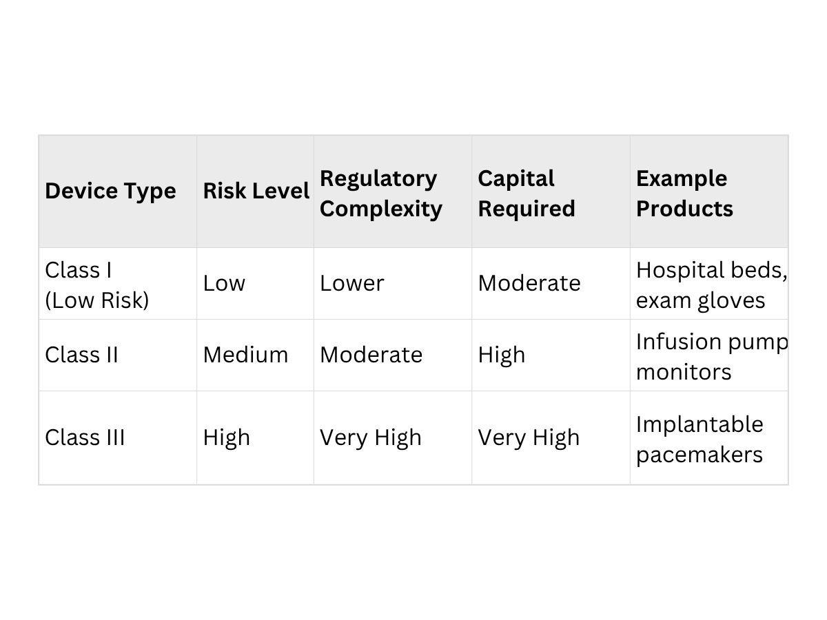
If you’re a first-time entrepreneur in this space, starting with lower-risk devices can reduce time to market and regulatory burden.
Building a Strong Foundation: Legal Structure
Forming a legal entity protects your personal assets and signals credibility to investors, partners, and regulators. Many entrepreneurs choose to register as a limited liability company (LLC) because it separates personal and business liability while maintaining operational flexibility.
Registering as an LLC can also simplify tax treatment and ownership structures, especially if you plan to bring on investors. You can reduce registration costs by self-filing with your state or using an online formation service. If you’re launching operations in California, you can explore options for setting up your LLC in California with ZenBusiness, which compares formation services and filing approaches.
The key is to formalize early. Manufacturers handling medical products should not operate informally.
A Practical Setup Checklist
Before you begin production, walk through this operational checklist:
1. Regulatory Planning
Determine device classification.
Identify required approvals or registrations.
2. Quality System Design
Implement documented manufacturing procedures.
Create traceability systems for materials and batches.
Train staff in quality control protocols.
3. Supplier Vetting
Audit suppliers for reliability and compliance.
Establish written quality agreements.
Avoid single-source dependency where possible.
4. Facility & Equipment
Ensure your manufacturing space meets safety and cleanliness standards.
Calibrate and validate production equipment.
5. Insurance Coverage
Secure product liability insurance.
Consider errors and omissions coverage if applicable.
Skipping any of these steps can create delays—or worse, product recalls.
Financing the Operation
Medical manufacturing often requires significant upfront investment. Tooling, molds, testing, and certifications are expensive.
Consider:
SBA loans or equipment financing.
Strategic partnerships with established distributors.
Angel or seed investment if developing innovative devices.
Pre-orders or pilot programs with local clinics.
Cash management matters more than flashy growth. Plan for longer sales cycles, especially when targeting hospitals.
Marketing & Distribution Strategy
Hospitals and clinics are cautious buyers. Trust is earned through:
Certifications and documented compliance.
Demonstrated product reliability.
Clear training materials and service agreements.
Responsive post-sale support.
Build relationships with:
Group purchasing organizations (GPOs).
Regional distributors.
Procurement officers in healthcare networks.
Reputation spreads quickly in healthcare—both good and bad.
Frequently Asked Questions
Do I need FDA approval for all medical devices?
Not always. Some low-risk devices require only registration and compliance with general controls, while others require premarket notification or approval. Classification determines the pathway.
How long does it take to launch?
For lower-risk devices, timelines may range from several months to over a year. Higher-risk devices can take multiple years due to testing and approval processes.
Is ISO certification mandatory?
Not always legally required, but many buyers expect compliance with recognized quality standards like ISO 13485.
Can I outsource manufacturing?
Yes, but you remain responsible for regulatory compliance and quality control. Oversight is critical.
A Helpful Industry Resource
For deeper insight into regulatory pathways and device classification, visit the U.S. Food and Drug Administration’s official medical device page. This resource provides up-to-date guidance on device categories, compliance expectations, and approval pathways. Always rely on official regulatory sources when making compliance decisions.
Common Early Mistakes to Avoid
Underestimating regulatory timelines
Neglecting documentation
Overbuilding before validating market demand
Choosing complex, high-risk products too early
Ignoring post-sale service planning
Entrepreneurs who thrive in this space treat quality, compliance, and customer trust as strategic assets—not overhead.
Starting a medical equipment manufacturing business requires discipline, capital, and patience. The opportunity is real—but so is the responsibility. Build slowly, document everything, and prioritize quality from day one. If you respect the regulatory environment and design your operations around compliance and reliability, you position your company for sustainable growth in a mission-critical industry.
Navigating regulatory hurdles, quality management, and market entry shouldn't hold back your medical innovation. Contact Metis Consulting Services to turn your healthcare vision into commercial reality.